 Français
 English

| ELK.No | ES10172 |
| Product name | RND1 rabbit pAb |
| Reactivity | Human;Mouse |
| Applications | WB;ELISA |
| Other name | |
| Size | 100μL |
| Unit price ($) | 248 |
| Human gene ID | 27289 |
| Human Swiss-Prot | Q92730 |
| Source | Rabbit |
| Isotype | IgG |
| Target | RND1 |
| Fields | >>Axon guidance |
| Gene name | RND1 RHO6 |
| Protein name | Rho-related GTP-binding protein Rho6 (Rho family GTPase 1) (Rnd1) |
| Human gene link | |
| Human Swiss link | View Human Swiss-Prot |
| Mouse gene ID | |
| Mouse gene link | |
| Mouse Swiss-Prot | Q8BLR7 |
| Mouse Swiss link | View Mouse Swiss-Prot |
| Rat gene ID | |
| Rat gene link | |
| Rat Swiss-Prot | |
| Rat Swiss link | |
| Immunogen | Synthesized peptide derived from human protein . at AA range: 140-220 |
| Specificity | RND1 Polyclonal Antibody detects endogenous levels of protein. |
| Formulation | Liquid in PBS containing 50% glycerol, and 0.02% sodium azide. |
| Clonality | Polyclonal |
| Dilution | WB 1:500-2000 ELISA 1:5000-20000 |
| Purification | The antibody was affinity-purified from rabbit antiserum by affinity-chromatography using epitope-specific immunogen. |
| Concentration | 1 mg/ml |
| Storage stability | -20°C/1 year |
| Molecular Weight (Da) | |
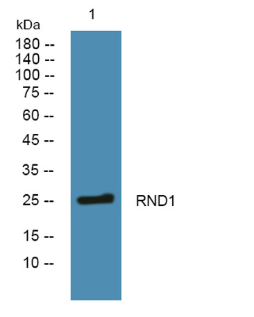
| Observed band (KD) | 25kD |
| Background | This gene encodes a protein that belongs to the Rho GTPase family. Members of this family regulate the organization of the actin cytoskeleton in response to extracellular growth factors. A similar protein in rat interacts with a microtubule regulator to control axon extension. [provided by RefSeq, Apr 2014], |
| Function | function:Lacks intrinsic GTPase activity. Has a low affinity for GDP, and constitutively binds GTP. Controls rearrangements of the actin cytoskeleton. Induces the Rac-dependent neuritic process formation in part by disruption of the cortical actin filaments. Causes the formation of many neuritic processes from the cell body with disruption of the cortical actin filaments.,similarity:Belongs to the small GTPase superfamily. Rho family.,subunit:Binds GRB7 and PLXNB1. Interacts with UBXD5.,tissue specificity:Mostly expressed in brain and liver., |
| Subcellular location | Cell membrane ; Lipid-anchor ; Cytoplasmic side . Cytoplasm, cytoskeleton . |
| Expression | Mostly expressed in brain and liver. |

Use collapsible tabs for more detailed information that will help customers make a purchasing decision.
Ex: Shipping and return policies, size guides, and other common questions.