 Français
 English

| ELK.No | ES10651 |
| Product name | FN3K rabbit pAb |
| Reactivity | Human;Mouse |
| Applications | WB;ELISA |
| Other name | |
| Size | 50μL |
| Unit price ($) | 148 |
| Human gene ID | 64122 |
| Human Swiss-Prot | Q9H479 |
| Source | Rabbit |
| Isotype | IgG |
| Target | FN3K |
| Fields | |
| Gene name | FN3K |
| Protein name | Fructosamine-3-kinase (EC 2.7.1.-) |
| Human gene link | |
| Human Swiss link | View Human Swiss-Prot |
| Mouse gene ID | |
| Mouse gene link | |
| Mouse Swiss-Prot | Q9ER35 |
| Mouse Swiss link | View Mouse Swiss-Prot |
| Rat gene ID | |
| Rat gene link | |
| Rat Swiss-Prot | |
| Rat Swiss link | |
| Immunogen | Synthesized peptide derived from human protein . at AA range: 130-210 |
| Specificity | FN3K Polyclonal Antibody detects endogenous levels of protein. |
| Formulation | Liquid in PBS containing 50% glycerol, and 0.02% sodium azide. |
| Clonality | Polyclonal |
| Dilution | WB 1:500-2000 ELISA 1:5000-20000 |
| Purification | The antibody was affinity-purified from rabbit antiserum by affinity-chromatography using epitope-specific immunogen. |
| Concentration | 1 mg/ml |
| Storage stability | -20°C/1 year |
| Molecular Weight (Da) | |
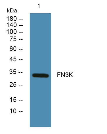
| Observed band (KD) | 33kD |
| Background | A high concentration of glucose can result in non-enzymatic oxidation of proteins by reaction of glucose and lysine residues (glycation). Proteins modified in this way, fructosamines, are less active or functional. This gene encodes an enzyme which catalyzes the phosphorylation of fructosamines which may result in deglycation. [provided by RefSeq, Feb 2012], |
| Function | function:May initiate a process leading to the deglycation of fructoselysine and of glycated proteins. May play a role in the phosphorylation of 1-deoxy-1-morpholinofructose (DMF), fructoselysine, fructoseglycine, fructose and glycated lysozyme.,similarity:Belongs to the fructosamine kinase family.,subunit:Monomer.,tissue specificity:Expressed in erythrocytes., |
| Subcellular location | cytosol, |
| Expression | Widely expressed (PubMed:11522682). Expressed in erythrocytes (PubMed:11016445). |

Use collapsible tabs for more detailed information that will help customers make a purchasing decision.
Ex: Shipping and return policies, size guides, and other common questions.