 Français
 English

| ELK.No | ES10676 |
| Product name | CCBE1 rabbit pAb |
| Reactivity | Human;Rat;Mouse; |
| Applications | WB;ELISA |
| Other name | |
| Size | 100μL |
| Unit price ($) | 248 |
| Human gene ID | 147372 |
| Human Swiss-Prot | Q6UXH8 |
| Source | Rabbit |
| Isotype | IgG |
| Target | CCBE1 |
| Fields | |
| Gene name | CCBE1 KIAA1983 UNQ1921/PRO4395 |
| Protein name | Collagen and calcium-binding EGF domain-containing protein 1 (Full of fluid protein homolog) |
| Human gene link | |
| Human Swiss link | View Human Swiss-Prot |
| Mouse gene ID | |
| Mouse gene link | |
| Mouse Swiss-Prot | Q3MI99 |
| Mouse Swiss link | View Mouse Swiss-Prot |
| Rat gene ID | |
| Rat gene link | |
| Rat Swiss-Prot | |
| Rat Swiss link | |
| Immunogen | Synthesized peptide derived from human protein . at AA range: 120-200 |
| Specificity | CCBE1 Polyclonal Antibody detects endogenous levels of protein. |
| Formulation | Liquid in PBS containing 50% glycerol, and 0.02% sodium azide. |
| Clonality | Polyclonal |
| Dilution | WB 1:500-2000 ELISA 1:5000-20000 |
| Purification | The antibody was affinity-purified from rabbit antiserum by affinity-chromatography using epitope-specific immunogen. |
| Concentration | 1 mg/ml |
| Storage stability | -20°C/1 year |
| Molecular Weight (Da) | |
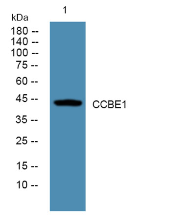
| Observed band (KD) | 44kD |
| Background | This gene is thought to function in extracellular matrix remodeling and migration. It is predominantly expressed in the ovary, but down regulated in ovarian cancer cell lines and primary carcinomas, suggesting its role as a tumour suppressor. Mutations in this gene have been associated with Hennekam lymphangiectasia-lymphedema syndrome, a generalized lymphatic dysplasia in humans. [provided by RefSeq, Mar 2010], |
| Function | function:Required for lymphangioblast budding and angiogenic sprouting from venous endothelium during embryogenesis.,similarity:Belongs to the CCBE1 family.,similarity:Contains 1 EGF-like domain.,similarity:Contains 2 collagen-like domains.,tissue specificity:Not expressed in blood or lymphatic endothelial cells., |
| Subcellular location | Secreted . |
| Expression | Not expressed in blood or lymphatic endothelial cells. |

Use collapsible tabs for more detailed information that will help customers make a purchasing decision.
Ex: Shipping and return policies, size guides, and other common questions.