 Français
 English
Ref-1 (Acetyl Lys7) rabbit pAb
AO-06-ES1098-100

| ELK.No | ES1098 |
| Product name | Ref-1 (Acetyl Lys7) rabbit pAb |
| Reactivity | Human;Rat;Mouse; |
| Applications | WB;IHC |
| Other name | APEX1; APE; APE1; APEX; APX; HAP1; REF1; DNA-(apurinic or apyrimidinic site) lyase; APEX nuclease; APEN; Apurinic-apyrimidinic endonuclease 1; AP endonuclease 1; APE-1; REF-1; Redox factor-1 |
| Size | 100μL |
| Unit price ($) | 248 |
| Human gene ID | 328 |
| Human Swiss-Prot | P27695 |
| Source | Rabbit |
| Isotype | IgG |
| Target | Ref-1 |
| Fields | >>Base excision repair |
| Gene name | APEX1 |
| Protein name | DNA-(apurinic or apyrimidinic site) lyase |
| Human gene link | View Human Gene |
| Human Swiss link | View Human Swiss-Prot |
| Mouse gene ID | |
| Mouse gene link | |
| Mouse Swiss-Prot | P28352 |
| Mouse Swiss link | View Mouse Swiss-Prot |
| Rat gene ID | |
| Rat gene link | |
| Rat Swiss-Prot | |
| Rat Swiss link | |
| Immunogen | The antiserum was produced against synthesized Acetyl-peptide derived from human APE1 around the Acetylation site of Lys7. AA range:1-50 |
| Specificity | Acetyl-Ref-1 (K7) Polyclonal Antibody detects endogenous levels of Ref-1 protein only when acetylated at K7. |
| Formulation | Liquid in PBS containing 50% glycerol, 0.5% BSA and 0.02% sodium azide. |
| Clonality | Polyclonal |
| Dilution | WB 1:500-2000;IHC-p 1:50-300 |
| Purification | The antibody was affinity-purified from rabbit antiserum by affinity-chromatography using epitope-specific immunogen. |
| Concentration | 1 mg/ml |
| Storage stability | -20°C/1 year |
| Molecular Weight (Da) | |
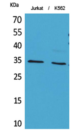
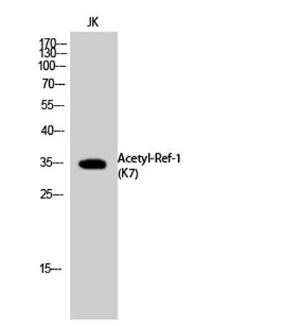
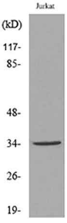
| Observed band (KD) | 35kD |
| Background | Apurinic/apyrimidinic (AP) sites occur frequently in DNA molecules by spontaneous hydrolysis, by DNA damaging agents or by DNA glycosylases that remove specific abnormal bases. AP sites are pre-mutagenic lesions that can prevent normal DNA replication so the cell contains systems to identify and repair such sites. Class II AP endonucleases cleave the phosphodiester backbone 5' to the AP site. This gene encodes the major AP endonuclease in human cells. Splice variants have been found for this gene; all encode the same protein. [provided by RefSeq, Jul 2008], |
| Function | catalytic activity:The C-O-P bond 3' to the apurinic or apyrimidinic site in DNA is broken by a beta-elimination reaction, leaving a 3'-terminal unsaturated sugar and a product with a terminal 5'-phosphate.,function:Repairs oxidative DNA damages in vitro. May have a role in protection against cell lethality and suppression of mutations. Removes the blocking groups from the 3'-termini of the DNA strand breaks generated by ionizing radiations and bleomycin.,similarity:Belongs to the DNA repair enzymes AP/exoA family.,subunit:Monomer. Component of the SET complex, which also contains SET, ANP32A, HMGB2 and NME1., |
| Subcellular location | Nucleus. Nucleus, nucleolus. Nucleus speckle. Endoplasmic reticulum. Cytoplasm. Detected in the cytoplasm of B-cells stimulated to switch (By similarity). Colocalized with SIRT1 in the nucleus. Colocalized with YBX1 in nuclear speckles after genotoxic stress. Together with OGG1 is recruited to nuclear speckles in UVA-irradiated cells. Colocalized with nucleolin and NPM1 in the nucleolus. Its nucleolar localization is cell cycle dependent and requires active rRNA transcription. Colocalized with calreticulin in the endoplasmic reticulum. Translocation from the nucleus to the cytoplasm is stimulated in presence of nitric oxide (NO) and function in a CRM1-dependent manner, possibly as a consequence of demasking a nuclear export signal (amino acid position 64-80). S-nitrosylation at Cys-93 and |
| Expression | Brain,Embryonic stem cells,Lung,Melanocyte,Placenta,Skin, |

Use collapsible tabs for more detailed information that will help customers make a purchasing decision.
Ex: Shipping and return policies, size guides, and other common questions.