 Français
 English

| ELK.No | ES11204 |
| Product name | HRK rabbit pAb |
| Reactivity | Human;Rat;Mouse |
| Applications | WB;ELISA |
| Other name | |
| Size | 100μL |
| Unit price ($) | 248 |
| Human gene ID | 8739 |
| Human Swiss-Prot | O00198 |
| Source | Rabbit |
| Isotype | IgG |
| Target | HRK |
| Fields | >>Apoptosis |
| Gene name | HRK BID3 |
| Protein name | Activator of apoptosis harakiri (BH3-interacting domain-containing protein 3) (Neuronal death protein DP5) |
| Human gene link | |
| Human Swiss link | View Human Swiss-Prot |
| Mouse gene ID | |
| Mouse gene link | |
| Mouse Swiss-Prot | P62816 |
| Mouse Swiss link | View Mouse Swiss-Prot |
| Rat gene ID | |
| Rat gene link | |
| Rat Swiss-Prot | P62817 |
| Rat Swiss link | View Rat Swiss-Prot |
| Immunogen | Synthesized peptide derived from part region of human protein AA range: 1-50 |
| Specificity | HRK Polyclonal Antibody detects endogenous levels of protein. |
| Formulation | Liquid in PBS containing 50% glycerol, and 0.02% sodium azide. |
| Clonality | Polyclonal |
| Dilution | WB 1:500-2000 ELISA 1:5000-20000 |
| Purification | The antibody was affinity-purified from rabbit antiserum by affinity-chromatography using epitope-specific immunogen. |
| Concentration | 1 mg/ml |
| Storage stability | -20°C/1 year |
| Molecular Weight (Da) | |
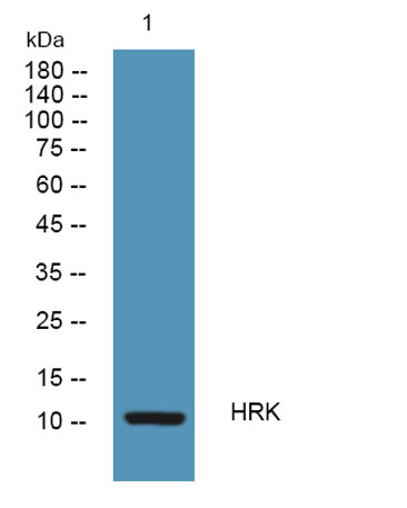
| Observed band (KD) | 10kD |
| Background | harakiri, BCL2 interacting protein(HRK) Homo sapiens This gene encodes a member of the BCL-2 protein family. Members of this family are involved in activating or inhibiting apoptosis. The encoded protein localizes to intracellular membranes. This protein promotes apoptosis by interacting with the apoptotic inhibitors BCL-2 and BCL-X(L) via its BH3 domain. Alternate splicing results in multiple transcript variants. [provided by RefSeq, Oct 2012], |
| Function | domain:The BH3 motif is required for the induction of cell death.,function:Activates apoptosis and interacts selectively with survival-promoting proteins Bcl-2 and Bcl-X(L)., |
| Subcellular location | Membrane; Single-pass membrane protein. Mitochondrion. |
| Expression |

Use collapsible tabs for more detailed information that will help customers make a purchasing decision.
Ex: Shipping and return policies, size guides, and other common questions.