 Français
 English

| ELK.No | ES11421 |
| Product name | SPXN rabbit pAb |
| Reactivity | Human;Rat;Mouse; |
| Applications | WB;ELISA |
| Other name | |
| Size | 100μL |
| Unit price ($) | 248 |
| Human gene ID | 80763 |
| Human Swiss-Prot | Q9BT56 |
| Source | Rabbit |
| Isotype | IgG |
| Target | SPXN |
| Fields | >>Neuroactive ligand-receptor interaction |
| Gene name | C12orf39 |
| Protein name | Spexin (NPQ) |
| Human gene link | |
| Human Swiss link | View Human Swiss-Prot |
| Mouse gene ID | |
| Mouse gene link | |
| Mouse Swiss-Prot | |
| Mouse Swiss link | |
| Rat gene ID | |
| Rat gene link | |
| Rat Swiss-Prot | |
| Rat Swiss link | |
| Immunogen | Synthesized peptide derived from human protein . at AA range: 1-50 |
| Specificity | SPXN Polyclonal Antibody detects endogenous levels of protein. |
| Formulation | Liquid in PBS containing 50% glycerol, and 0.02% sodium azide. |
| Clonality | Polyclonal |
| Dilution | WB 1:500-2000 ELISA 1:5000-20000 |
| Purification | The antibody was affinity-purified from rabbit antiserum by affinity-chromatography using epitope-specific immunogen. |
| Concentration | 1 mg/ml |
| Storage stability | -20°C/1 year |
| Molecular Weight (Da) | |
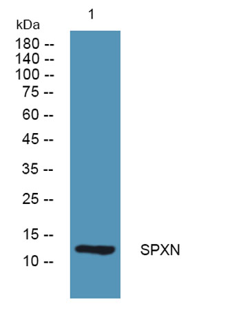
| Observed band (KD) | 12kD |
| Background | The protein encoded by this gene is a hormone involved in modulation of cardiovascular and renal function. It has also been shown in rats to cause weight loss. Several transcript variants have been found for this gene. [provided by RefSeq, Feb 2016], |
| Function | function:Induces contraction of stomach muscle.,tissue specificity:Expressed in submucosal layer of esophagus and stomach fundus. Expressed in brain, pancreas and kidney., |
| Subcellular location | Secreted. Secreted, extracellular space. Cytoplasmic vesicle, secretory vesicle. Secreted via the classical ER/Golgi-dependent pathway into the extracellular medium largely as a full-length protein without the signal peptide, and not as a hydrolyzed and amidated peptide (PubMed:19193193 and PubMed:17284679). Localized extracellularly surrounding the villous trophoblastic cells. Detected in the serum. |
| Expression | Expressed in the type I glomic cells within the carotid body (at protein level). Expressed predominantly in pancreas, testis, kidney, brain and placenta. Expressed in submucosal layer of esophagus and stomach fundus. |

Use collapsible tabs for more detailed information that will help customers make a purchasing decision.
Ex: Shipping and return policies, size guides, and other common questions.