 Français
 English

| ELK.No | ES12697 |
| Product name | TIGD7 rabbit pAb |
| Reactivity | Human;Rat;Mouse; |
| Applications | WB;IHC |
| Other name | |
| Size | 100μL |
| Unit price ($) | 248 |
| Human gene ID | 91151 |
| Human Swiss-Prot | Q6NT04 |
| Source | Rabbit |
| Isotype | IgG |
| Target | TIGD7 |
| Fields | |
| Gene name | TIGD7 |
| Protein name | TIGD7 |
| Human gene link | View Human Gene |
| Human Swiss link | View Human Swiss-Prot |
| Mouse gene ID | |
| Mouse gene link | |
| Mouse Swiss-Prot | |
| Mouse Swiss link | |
| Rat gene ID | |
| Rat gene link | |
| Rat Swiss-Prot | |
| Rat Swiss link | |
| Immunogen | Synthesized peptide derived from human TIGD7 AA range: 308-358 |
| Specificity | This antibody detects endogenous levels of TIGD7 at Human |
| Formulation | Liquid in PBS containing 50% glycerol, 0.5% BSA and 0.02% sodium azide. |
| Clonality | Polyclonal |
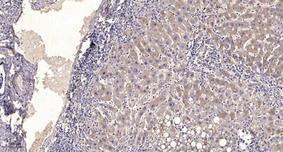
| Dilution | WB 1:500-2000;IHC-p 1:50-300 |
| Purification | The antibody was affinity-purified from rabbit serum by affinity-chromatography using specific immunogen. |
| Concentration | 1 mg/ml |
| Storage stability | -20°C/1 year |
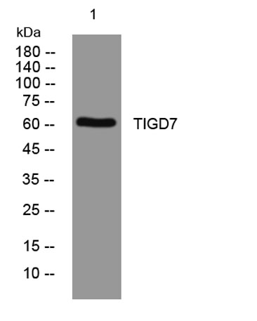
| Molecular Weight (Da) | 60kD |
| Observed band (KD) | |
| Background | The protein encoded by this gene belongs to the tigger subfamily of the pogo superfamily of DNA-mediated transposons in humans. These proteins are related to DNA transposons found in fungi and nematodes, and more distantly to the Tc1 and mariner transposases. They are also very similar to the major mammalian centromere protein B. The exact function of this gene is not known. [provided by RefSeq, Jul 2008], |
| Function | similarity:Belongs to the tigger transposable element derived protein family.,similarity:Contains 1 DDE domain.,similarity:Contains 1 HTH CENPB-type DNA-binding domain.,similarity:Contains 1 HTH psq-type DNA-binding domain.,tissue specificity:Expressed in all tissues tested. Higher expression in testis and ovary., |
| Subcellular location | Nucleus . |
| Expression | Expressed in all tissues tested. Higher expression in testis and ovary. |

Use collapsible tabs for more detailed information that will help customers make a purchasing decision.
Ex: Shipping and return policies, size guides, and other common questions.