 Français
 English

| ELK.No | ES12796 |
| Product name | TBCD7 rabbit pAb |
| Reactivity | Human; Mouse |
| Applications | WB |
| Other name | |
| Size | 100μL |
| Unit price ($) | 248 |
| Human gene ID | 51256 |
| Human Swiss-Prot | Q9P0N9 |
| Source | Rabbit |
| Isotype | IgG |
| Target | TBCD7 |
| Fields | >>mTOR signaling pathway |
| Gene name | TBC1D7 HSPC239 TBC7 |
| Protein name | TBCD7 |
| Human gene link | View Human Gene |
| Human Swiss link | View Human Swiss-Prot |
| Mouse gene ID | 67046 |
| Mouse gene link | View Mouse Gene |
| Mouse Swiss-Prot | Q9D0K0 |
| Mouse Swiss link | View Mouse Swiss-Prot |
| Rat gene ID | |
| Rat gene link | |
| Rat Swiss-Prot | |
| Rat Swiss link | |
| Immunogen | Synthesized peptide derived from human TBCD7 AA range: 85-135 |
| Specificity | This antibody detects endogenous levels of TBCD7 at Human/Mouse |
| Formulation | Liquid in PBS containing 50% glycerol, 0.5% BSA and 0.02% sodium azide. |
| Clonality | Polyclonal |
| Dilution | WB 1:500-2000 |
| Purification | The antibody was affinity-purified from rabbit serum by affinity-chromatography using specific immunogen. |
| Concentration | 1 mg/ml |
| Storage stability | -20°C/1 year |
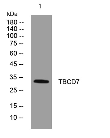
| Molecular Weight (Da) | 32kD |
| Observed band (KD) | |
| Background | This gene encodes a member of the TBC-domain containing protein family. The encoded protein functions as a subunit of the tuberous sclerosis TSC1-TSC2 complex which plays a role in the regulation of cellular growth and differentiation. Mutations in this gene have been associated with autosomal recessive megalencephaly. Alternative splicing results in multiple transcript variants. Naturally occurring readthrough transcription occurs between this locus and downstream LOC100130357. [provided by RefSeq, Jan 2016], |
| Function | function:May act as a GTPase-activating protein for Rab family protein(s) .,similarity:Contains 1 Rab-GAP TBC domain., |
| Subcellular location | Cytoplasmic vesicle . Localizes in the cytoplasmic vesicles of the endomembrane in association with TSC1-TSC2 complex. |
| Expression | Highly expressed in heart, and slightly in kidney, liver and placenta. |

Use collapsible tabs for more detailed information that will help customers make a purchasing decision.
Ex: Shipping and return policies, size guides, and other common questions.