 Français
 English
Chk2 (phospho Thr68) rabbit pAb
AO-06-ES1288-50

| ELK.No | ES1288 |
| Product name | Chk2 (phospho Thr68) rabbit pAb |
| Reactivity | Human;Mouse;Rat |
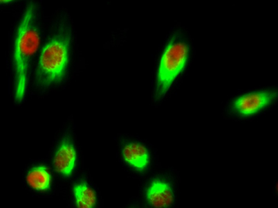
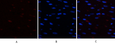
| Applications | IF;WB;IHC;ELISA |
| Other name | CHEK2; CDS1; CHK2; RAD53; Serine/threonine-protein kinase Chk2; CHK2 checkpoint homolog; Cds1 homolog; Hucds1; hCds1; Checkpoint kinase 2 |
| Size | 50μL |
| Unit price ($) | 148 |
| Human gene ID | 11200 |
| Human Swiss-Prot | O96017 |
| Source | Rabbit |
| Isotype | IgG |
| Target | Chk2 |
| Fields | >>Cell cycle;>>p53 signaling pathway;>>Cellular senescence;>>Human T-cell leukemia virus 1 infection |
| Gene name | CHEK2 |
| Protein name | Serine/threonine-protein kinase Chk2 |
| Human gene link | View Human Gene |
| Human Swiss link | View Human Swiss-Prot |
| Mouse gene ID | 50883 |
| Mouse gene link | View Mouse Gene |
| Mouse Swiss-Prot | Q9Z265 |
| Mouse Swiss link | View Mouse Swiss-Prot |
| Rat gene ID | |
| Rat gene link | |
| Rat Swiss-Prot | |
| Rat Swiss link | |
| Immunogen | The antiserum was produced against synthesized peptide derived from human Chk2 around the phosphorylation site of Thr68. AA range:35-84 |
| Specificity | Phospho-Chk2 (T68) Polyclonal Antibody detects endogenous levels of Chk2 protein only when phosphorylated at T68. |
| Formulation | Liquid in PBS containing 50% glycerol, 0.5% BSA and 0.02% sodium azide. |
| Clonality | Polyclonal |
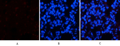
| Dilution | IF: 1:50-200 Western Blot: 1/500 - 1/2000. Immunohistochemistry: 1/100 - 1/300. ELISA: 1/20000. Not yet tested in other applications. |
| Purification | The antibody was affinity-purified from rabbit antiserum by affinity-chromatography using epitope-specific immunogen. |
| Concentration | 1 mg/ml |
| Storage stability | -20°C/1 year |
| Molecular Weight (Da) | |
| Observed band (KD) | 61kD |
| Background | In response to DNA damage and replication blocks, cell cycle progression is halted through the control of critical cell cycle regulators. The protein encoded by this gene is a cell cycle checkpoint regulator and putative tumor suppressor. It contains a forkhead-associated protein interaction domain essential for activation in response to DNA damage and is rapidly phosphorylated in response to replication blocks and DNA damage. When activated, the encoded protein is known to inhibit CDC25C phosphatase, preventing entry into mitosis, and has been shown to stabilize the tumor suppressor protein p53, leading to cell cycle arrest in G1. In addition, this protein interacts with and phosphorylates BRCA1, allowing BRCA1 to restore survival after DNA damage. Mutations in this gene have been linked with Li-Fraumeni syndrome, a highly penetrant familial cancer phenotype usually associated with inherited mutati |
| Function | catalytic activity:ATP + a protein = ADP + a phosphoprotein.,cofactor:Magnesium.,disease:Defects in CHEK2 are associated with Li-Fraumeni syndrome 2 (LFS2) [MIM:609265]; a highly penetrant familial cancer phenotype usually associated with inherited mutations in p53/TP53.,disease:Defects in CHEK2 are found in some patients with osteosarcoma (OSRC) [MIM:259500].,disease:Defects in CHEK2 are found in some patients with prostate cancer (CaP) [MIM:176807].,enzyme regulation:Rapidly phosphorylated on Thr-68 by MLTK in response to DNA damage and to replication block. Kinase activity is also up-regulated by autophosphorylation.,function:Regulates cell cycle checkpoints and apoptosis in response to DNA damage, particularly to DNA double-strand breaks. Inhibits CDC25C phosphatase by phosphorylation on 'Ser-216', preventing the entry into mitosis. May also play a role in meiosis. Regulates the TP53 |
| Subcellular location | [Isoform 2]: Nucleus. Isoform 10 is present throughout the cell.; [Isoform 4]: Nucleus.; [Isoform 7]: Nucleus.; [Isoform 9]: Nucleus.; [Isoform 12]: Nucleus.; Nucleus, PML body. Nucleus, nucleoplasm. Recruited into PML bodies together with TP53. |
| Expression | High expression is found in testis, spleen, colon and peripheral blood leukocytes. Low expression is found in other tissues. |

Use collapsible tabs for more detailed information that will help customers make a purchasing decision.
Ex: Shipping and return policies, size guides, and other common questions.