 Français
 English

| ELK.No | ES12903 |
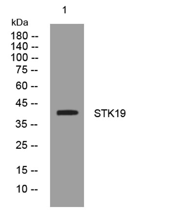
| Product name | STK19 rabbit pAb |
| Reactivity | Human; Mouse |
| Applications | WB |
| Other name | |
| Size | 50μL |
| Unit price ($) | 148 |
| Human gene ID | 8859 |
| Human Swiss-Prot | P49842 |
| Source | Rabbit |
| Isotype | IgG |
| Target | STK19 |
| Fields | |
| Gene name | STK19 G11 RP1 |
| Protein name | STK19 |
| Human gene link | View Human Gene |
| Human Swiss link | View Human Swiss-Prot |
| Mouse gene ID | 54402 |
| Mouse gene link | View Mouse Gene |
| Mouse Swiss-Prot | Q9JHN8 |
| Mouse Swiss link | View Mouse Swiss-Prot |
| Rat gene ID | |
| Rat gene link | |
| Rat Swiss-Prot | |
| Rat Swiss link | |
| Immunogen | Synthesized peptide derived from human STK19 AA range: 89-139 |
| Specificity | This antibody detects endogenous levels of STK19 at Human/Mouse |
| Formulation | Liquid in PBS containing 50% glycerol, 0.5% BSA and 0.02% sodium azide. |
| Clonality | Polyclonal |
| Dilution | WB 1:500-2000 |
| Purification | The antibody was affinity-purified from rabbit serum by affinity-chromatography using specific immunogen. |
| Concentration | 1 mg/ml |
| Storage stability | -20°C/1 year |
| Molecular Weight (Da) | 40kD |
| Observed band (KD) | |
| Background | This gene encodes a serine/threonine kinase which localizes predominantly to the nucleus. Its specific function is unknown; it is possible that phosphorylation of this protein is involved in transcriptional regulation. This gene localizes to the major histocompatibility complex (MHC) class III region on chromosome 6 and expresses two transcript variants. [provided by RefSeq, Jul 2008], |
| Function | catalytic activity:ATP + a protein = ADP + a phosphoprotein.,cofactor:Divalent cations. Has a preference for manganese.,function:Seems to be a protein kinase. In vitro it can phosphorylate casein-alpha on serine and threonine residues and histones on serine residues.,similarity:Belongs to the STK19 family.,subunit:The N-terminus interacts with BAG1.,tissue specificity:Monocytes, hepatocytes, epithelial cells, T-and B-lymphocytes., |
| Subcellular location | Nucleus . |
| Expression | Monocytes, hepatocytes, epithelial cells, T- and B-lymphocytes. |

Use collapsible tabs for more detailed information that will help customers make a purchasing decision.
Ex: Shipping and return policies, size guides, and other common questions.