 Français
 English

| ELK.No | ES12993 |
| Product name | SPEB rabbit pAb |
| Reactivity | Human; Mouse;Rat |
| Applications | WB |
| Other name | Agmatinase, mitochondrial (EC 3.5.3.11) (Agmatine ureohydrolase) (AUH) |
| Size | 50μL |
| Unit price ($) | 148 |
| Human gene ID | 79814 |
| Human Swiss-Prot | Q9BSE5 |
| Source | Rabbit |
| Isotype | IgG |
| Target | SPEB |
| Fields | >>Arginine and proline metabolism;>>Metabolic pathways |
| Gene name | AGMAT |
| Protein name | SPEB |
| Human gene link | View Human Gene |
| Human Swiss link | View Human Swiss-Prot |
| Mouse gene ID | 75986 |
| Mouse gene link | View Mouse Gene |
| Mouse Swiss-Prot | A2AS89 |
| Mouse Swiss link | View Mouse Swiss-Prot |
| Rat gene ID | 298607 |
| Rat gene link | View Rat Gene |
| Rat Swiss-Prot | Q0D2L3 |
| Rat Swiss link | View Rat Swiss-Prot |
| Immunogen | Synthesized peptide derived from human SPEB AA range: 266-316 |
| Specificity | This antibody detects endogenous levels of SPEB at Human/Mouse/Rat |
| Formulation | Liquid in PBS containing 50% glycerol, 0.5% BSA and 0.25% sodium azide. |
| Clonality | Polyclonal |
| Dilution | WB 1:500-2000 |
| Purification | The antibody was affinity-purified from rabbit serum by affinity-chromatography using specific immunogen. |
| Concentration | 1 mg/ml |
| Storage stability | -20°C/1 year |
| Molecular Weight (Da) | |
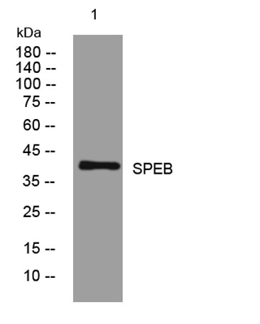
| Observed band (KD) | 38kD |
| Background | |
| Function | catalytic activity:Agmatine + H(2)O = putrescine + urea.,cofactor:Manganese .,pathway:Amine and polyamine biosynthesis; putrescine biosynthesis via agmatine pathway; putrescine from agmatine (ADC route): step 1/1.,similarity:Belongs to the arginase family. Agmatinase subfamily.,tissue specificity:Highly expressed in liver and kidney. Also found in skeletal muscle, fetal liver, brain, testis, skin and the gastrointestinal tract., |
| Subcellular location | Mitochondrion. |
| Expression | Highly expressed in liver and kidney. Also found in skeletal muscle, fetal liver, brain, testis, skin and the gastrointestinal tract. Within brain, expression is higher in the cerebral cortex with lower levels in the medulla and spinal cord. |

Use collapsible tabs for more detailed information that will help customers make a purchasing decision.
Ex: Shipping and return policies, size guides, and other common questions.