 Français
 English
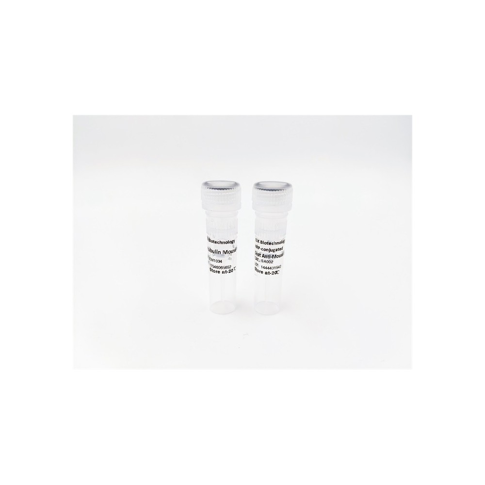

| ELK.No | ES13378 |
| Product name | RMXL1 rabbit pAb |
| Reactivity | Human; Mouse;Rat |
| Applications | WB;ELISA;IHC |
| Other name | |
| Size | 50μL |
| Unit price ($) | 148 |
| Human gene ID | 494115 |
| Human Swiss-Prot | Q96E39 |
| Source | Rabbit |
| Isotype | IgG |
| Target | RMXL1 |
| Fields | |
| Gene name | RBMXL1 |
| Protein name | RMXL1 |
| Human gene link | View Human Gene |
| Human Swiss link | View Human Swiss-Prot |
| Mouse gene ID | 19656 |
| Mouse gene link | View Mouse Gene |
| Mouse Swiss-Prot | Q91VM5 |
| Mouse Swiss link | View Mouse Swiss-Prot |
| Rat gene ID | |
| Rat gene link | |
| Rat Swiss-Prot | D4AE41 |
| Rat Swiss link | View Rat Swiss-Prot |
| Immunogen | Synthesized peptide derived from human RMXL1 AA range: 304-354 |
| Specificity | This antibody detects endogenous levels of RMXL1 at Human/Mouse/Rat |
| Formulation | Liquid in PBS containing 50% glycerol, 0.5% BSA and 0.02% sodium azide. |
| Clonality | Polyclonal |
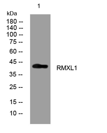
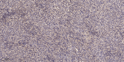
| Dilution | WB 1:500-2000;IHC-p 1:50-300; ELISA 2000-20000 |
| Purification | The antibody was affinity-purified from rabbit serum by affinity-chromatography using specific immunogen. |
| Concentration | 1 mg/ml |
| Storage stability | -20°C/1 year |
| Molecular Weight (Da) | 43kD |
| Observed band (KD) | |
| Background | This gene represents a retrogene of RNA binding motif protein, X-linked (RBMX), which is located on chromosome X. While all introns in the coding sequence have been processed out compared to the RBMX locus, the ORF is intact and there is specific evidence for transcription at this location. The preservation of the ORF by purifying selection in all Old World monkeys carrying it suggests that this locus is likely to be functional, possibly during male meiosis when X chromosomal genes are silenced or during haploid stages of spermatogenesis. This gene shares 5' exon structure with the cysteine conjugate-beta lyase 2 locus on chromosome 1, but the coding sequences are non-overlapping. Alternative splicing results in two transcript variants. [provided by RefSeq, Jun 2009], |
| Function | |
| Subcellular location | Nucleus . |
| Expression |

Use collapsible tabs for more detailed information that will help customers make a purchasing decision.
Ex: Shipping and return policies, size guides, and other common questions.