 Français
 English
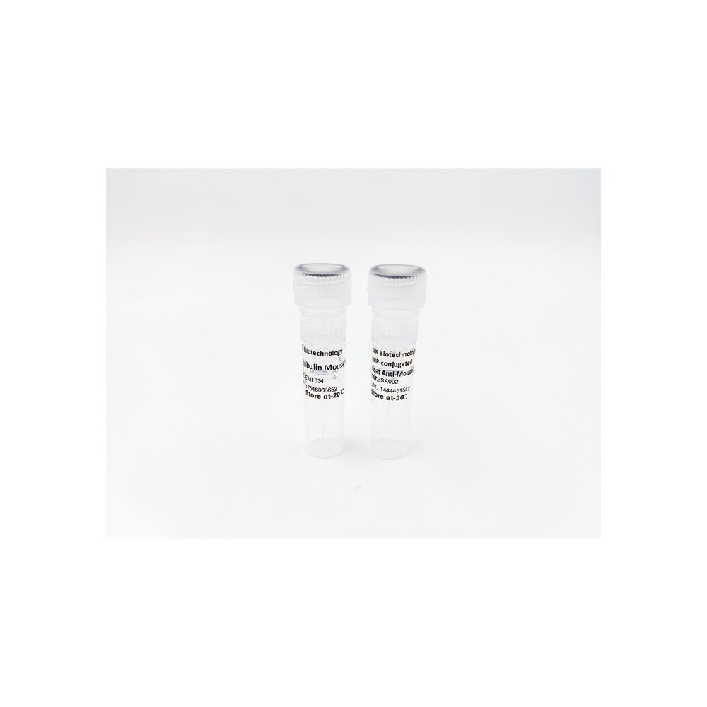

| ELK.No | ES13447 |
| Product name | RBM25 rabbit pAb |
| Reactivity | Human; Mouse |
| Applications | WB;IHC |
| Other name | |
| Size | 50μL |
| Unit price ($) | 148 |
| Human gene ID | 58517 |
| Human Swiss-Prot | P49756 |
| Source | Rabbit |
| Isotype | IgG |
| Target | RBM25 |
| Fields | >>Spliceosome |
| Gene name | RBM25 RNPC7 |
| Protein name | RBM25 |
| Human gene link | View Human Gene |
| Human Swiss link | View Human Swiss-Prot |
| Mouse gene ID | 67039 |
| Mouse gene link | View Mouse Gene |
| Mouse Swiss-Prot | B2RY56 |
| Mouse Swiss link | View Mouse Swiss-Prot |
| Rat gene ID | |
| Rat gene link | |
| Rat Swiss-Prot | |
| Rat Swiss link | |
| Immunogen | Synthesized peptide derived from human RBM25 AA range: 763-813 |
| Specificity | This antibody detects endogenous levels of RBM25 at Human/Mouse |
| Formulation | Liquid in PBS containing 50% glycerol, 0.5% BSA and 0.02% sodium azide. |
| Clonality | Polyclonal |
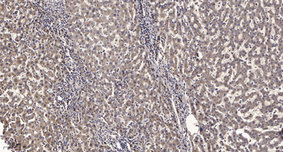
| Dilution | WB 1:500-2000;IHC-p 1:50-300 |
| Purification | The antibody was affinity-purified from rabbit serum by affinity-chromatography using specific immunogen. |
| Concentration | 1 mg/ml |
| Storage stability | -20°C/1 year |
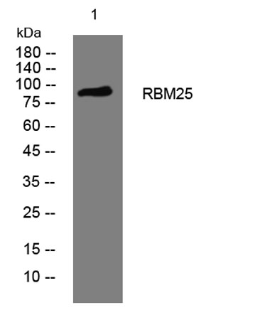
| Molecular Weight (Da) | 93kD |
| Observed band (KD) | |
| Background | |
| Function | PTM:Sumoylated.,sequence caution:Contaminating sequence. Potential poly-A sequence.,sequence caution:Translation N-terminally extended.,similarity:Contains 1 PWI domain.,similarity:Contains 1 RRM (RNA recognition motif) domain.,subcellular location:Detected predominantly in nuclear speckles. Cytoplasmic localization is faint.,subunit:Interacts with SRRM1. Specifically associates with functional splicing complexes, including Sm proteins and U1, U2, U4, U5 and U6 snRNAs. Associates with exon junction complex (EJC) proteins, including APEX1, BAT1, NCBP1, RBM8A and RNPS1. Interaction with NCBP1 is RNA-dependent., |
| Subcellular location | Nucleus speckle. Cytoplasm. Colocalizes predominantly, with SFRS2 and LUC7L3 splicing factors, in nuclear speckles. Cytoplasmic localization is faint. |
| Expression |

Use collapsible tabs for more detailed information that will help customers make a purchasing decision.
Ex: Shipping and return policies, size guides, and other common questions.