 Français
 English
Progesterone Receptor (phospho-Ser345) rabbit pAb
AO-06-ES13943-100

| ELK.No | ES13943 |
| Product name | Progesterone Receptor (phospho-Ser345) rabbit pAb |
| Reactivity | Human;Rat;Mouse; |
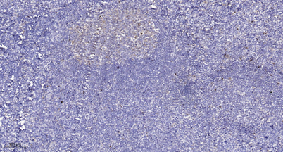
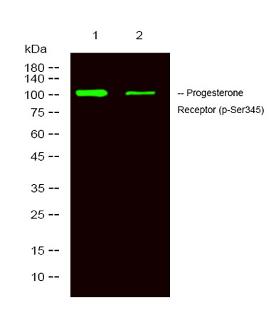
| Applications | WB;ELISA;IHC |
| Other name | Progesterone receptor (PR) (Nuclear receptor subfamily 3 group C member 3) |
| Size | 100μL |
| Unit price ($) | 248 |
| Human gene ID | 5241 |
| Human Swiss-Prot | P06401 |
| Source | Rabbit |
| Isotype | IgG |
| Target | PR |
| Fields | >>Oocyte meiosis;>>Progesterone-mediated oocyte maturation;>>Estrogen signaling pathway;>>Chemical carcinogenesis - receptor activation;>>Breast cancer |
| Gene name | PGR NR3C3 |
| Protein name | Progesterone Receptor (Ser345) |
| Human gene link | View Human Gene |
| Human Swiss link | View Human Swiss-Prot |
| Mouse gene ID | |
| Mouse gene link | |
| Mouse Swiss-Prot | Q00175 |
| Mouse Swiss link | View Mouse Swiss-Prot |
| Rat gene ID | 25154 |
| Rat gene link | View Rat Gene |
| Rat Swiss-Prot | Q63449 |
| Rat Swiss link | View Rat Swiss-Prot |
| Immunogen | Synthesized phosho peptide around human Progesterone Receptor (Ser345) |
| Specificity | This antibody detects endogenous levels of Human Progesterone Receptor (phospho-Ser345) |
| Formulation | Liquid in PBS containing 50% glycerol, 0.5% BSA and 0.02% sodium azide. |
| Clonality | Polyclonal |
| Dilution | WB 1:500-2000;IHC-p 1:50-300; ELISA 2000-20000 |
| Purification | The antibody was affinity-purified from rabbit serum by affinity-chromatography using specific immunogen. |
| Concentration | 1 mg/ml |
| Storage stability | -20°C/1 year |
| Molecular Weight (Da) | |
| Observed band (KD) | 105kD |
| Background | This gene encodes a member of the steroid receptor superfamily. The encoded protein mediates the physiological effects of progesterone, which plays a central role in reproductive events associated with the establishment and maintenance of pregnancy. This gene uses two distinct promotors and translation start sites in the first exon to produce several transcript variants, both protein coding and non-protein coding. Two of the isoforms (A and B) are identical except for an additional 165 amino acids found in the N-terminus of isoform B and mediate their own response genes and physiologic effects with little overlap. [provided by RefSeq, Sep 2015], |
| Function | domain:Composed of three domains: a modulating N-terminal domain, a DNA-binding domain and a C-terminal steroid-binding domain.,function:Isoform A is inactive in stimulating c-Src/MAPK signaling on hormone stimulation.,function:The steroid hormones and their receptors are involved in the regulation of eukaryotic gene expression and affect cellular proliferation and differentiation in target tissues. Progesterone receptor isoform B (PRB) is involved activation of c-SRC/MAPK signaling on hormone stimulation.,online information:Progesterone receptor entry,PTM:Phosphorylated on multiple serine sites. Several of these sites are hormone-dependent. Phosphorylation on Ser-294 occurs preferentially on isoform B, is highly hormone-dependent and modulates ubiquitination and sumoylation on Lys-388. Phosphorylation on Ser-102 and Ser-345 also requires induction by hormone. Basal phosphorylation on Se |
| Subcellular location | Nucleus. Cytoplasm. Nucleoplasmic shuttling is both hormone- and cell cycle-dependent. On hormone stimulation, retained in the cytoplasm in the G(1) and G(2)/M phases.; [Isoform A]: Nucleus. Cytoplasm. Mainly nuclear.; [Isoform 4]: Mitochondrion outer membrane . |
| Expression | In reproductive tissues the expression of isoform A and isoform B varies as a consequence of developmental and hormonal status. Isoform A and isoform B are expressed in comparable levels in uterine glandular epithelium during the proliferative phase of the menstrual cycle. Expression of isoform B but not of isoform A persists in the glands during mid-secretory phase. In the stroma, isoform A is the predominant form throughout the cycle. Heterogeneous isoform expression between the glands of the endometrium basalis and functionalis is implying region-specific responses to hormonal stimuli. |

Use collapsible tabs for more detailed information that will help customers make a purchasing decision.
Ex: Shipping and return policies, size guides, and other common questions.