 Français
 English
VASP (phospho Ser157) rabbit pAb
AO-06-ES1414-50

| ELK.No | ES1414 |
| Product name | VASP (phospho Ser157) rabbit pAb |
| Reactivity | Human;Mouse;Rat;Monkey |
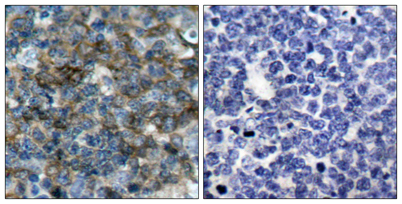
| Applications | WB;IHC;IF;ELISA |
| Other name | VASP; Vasodilator-stimulated phosphoprotein; VASP |
| Size | 50μL |
| Unit price ($) | 148 |
| Human gene ID | 7408 |
| Human Swiss-Prot | P50552 |
| Source | Rabbit |
| Isotype | IgG |
| Target | VASP |
| Fields | >>Rap1 signaling pathway;>>cGMP-PKG signaling pathway;>>Focal adhesion;>>Tight junction;>>Platelet activation;>>Fc gamma R-mediated phagocytosis;>>Leukocyte transendothelial migration |
| Gene name | VASP |
| Protein name | Vasodilator-stimulated phosphoprotein |
| Human gene link | View Human Gene |
| Human Swiss link | View Human Swiss-Prot |
| Mouse gene ID | 22323 |
| Mouse gene link | View Mouse Gene |
| Mouse Swiss-Prot | P70460 |
| Mouse Swiss link | View Mouse Swiss-Prot |
| Rat gene ID | |
| Rat gene link | |
| Rat Swiss-Prot | |
| Rat Swiss link | |
| Immunogen | The antiserum was produced against synthesized peptide derived from human VASP around the phosphorylation site of Ser157. AA range:124-173 |
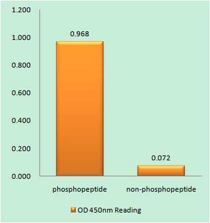
| Specificity | Phospho-VASP (S157) Polyclonal Antibody detects endogenous levels of VASP protein only when phosphorylated at S157. |
| Formulation | Liquid in PBS containing 50% glycerol, 0.5% BSA and 0.02% sodium azide. |
| Clonality | Polyclonal |
| Dilution | Western Blot: 1/500 - 1/2000. Immunohistochemistry: 1/100 - 1/300. ELISA: 1/5000. Not yet tested in other applications. |
| Purification | The antibody was affinity-purified from rabbit antiserum by affinity-chromatography using epitope-specific immunogen. |
| Concentration | 1 mg/ml |
| Storage stability | -20°C/1 year |
| Molecular Weight (Da) | |
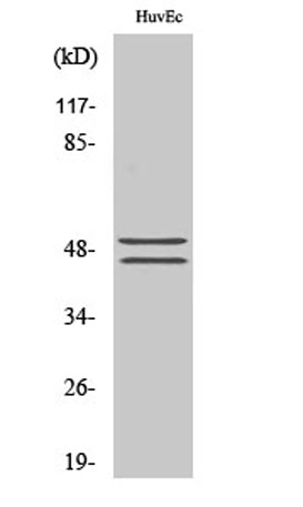
| Observed band (KD) | 46+50kD |
| Background | Vasodilator-stimulated phosphoprotein (VASP) is a member of the Ena-VASP protein family. Ena-VASP family members contain an EHV1 N-terminal domain that binds proteins containing E/DFPPPPXD/E motifs and targets Ena-VASP proteins to focal adhesions. In the mid-region of the protein, family members have a proline-rich domain that binds SH3 and WW domain-containing proteins. Their C-terminal EVH2 domain mediates tetramerization and binds both G and F actin. VASP is associated with filamentous actin formation and likely plays a widespread role in cell adhesion and motility. VASP may also be involved in the intracellular signaling pathways that regulate integrin-extracellular matrix interactions. VASP is regulated by the cyclic nucleotide-dependent kinases PKA and PKG. [provided by RefSeq, Jul 2008], |
| Function | domain:The EVH2 domain is comprised of 3 regions. Block A is a thymosin-like domain required for G-actin binding. The KLKR motif within this block is essential for the G-actin binding and for actin polymerization. Block B is required for F-actin binding and subcellular location, and Block C for tetramerization.,domain:The WH1 domain mediates interaction with XIRP1.,function:Ena/VASP proteins are actin-associated proteins involved in a range of processes dependent on cytoskeleton remodeling and cell polarity such as axon guidance and lamellipodial and filopodial dynamics in migrating cells. VASP promotes actin nucleation and increases the rate of actin polymerization in the presence of capping protein. Plays a role in actin-based activity of Listeria monocytogenes in platelets.,PTM:Major substrate for cAMP-dependent (PKA) and cGMP-dependent protein kinase (PKG) in platelets. The preferred |
| Subcellular location | Cytoplasm. Cytoplasm, cytoskeleton. Cell junction, focal adhesion. Cell junction, tight junction . Cell projection, lamellipodium membrane. Cell projection, filopodium membrane. Targeted to stress fibers and focal adhesions through interaction with a number of proteins including MRL family members. Localizes to the plasma membrane in protruding lamellipodia and filopodial tips. Stimulation by thrombin or PMA, also translocates VASP to focal adhesions. Localized along the sides of actin filaments throughout the peripheral cytoplasm under basal conditions. In pre-apoptotic cells, colocalizes with MEFV in large specks (pyroptosomes). |
| Expression | Highly expressed in platelets. |

Use collapsible tabs for more detailed information that will help customers make a purchasing decision.
Ex: Shipping and return policies, size guides, and other common questions.