 Français
 English

PDGF Receptor α (phospho-Tyr1018) rabbit pAb
AO-06-ES14184-100

| ELK.No | ES14184 |
| Product name | PDGF Receptor α (phospho-Tyr1018) rabbit pAb |
| Reactivity | Human;Mouse |
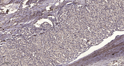
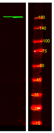
| Applications | WB;ELISA;IHC |
| Other name | Platelet-derived growth factor receptor alpha (PDGF-R-alpha) (PDGFR-alpha) (EC 2.7.10.1) (Alpha platelet-derived growth factor receptor) (Alpha-type platelet-derived growth factor receptor) (CD140 antigen-like family member A) (CD140a antigen) (Platelet-derived growth factor alpha receptor) (Platelet-derived growth factor receptor 2) (PDGFR-2) (CD antigen CD140a) |
| Size | 100μL |
| Unit price ($) | 248 |
| Human gene ID | 5156 |
| Human Swiss-Prot | P16234 |
| Source | Rabbit |
| Isotype | IgG |
| Target | PDGF Receptor α |
| Fields | >>EGFR tyrosine kinase inhibitor resistance;>>MAPK signaling pathway;>>Ras signaling pathway;>>Rap1 signaling pathway;>>Calcium signaling pathway;>>Phospholipase D signaling pathway;>>Endocytosis;>>PI3K-Akt signaling pathway;>>Focal adhesion;>>Gap junction;>>JAK-STAT signaling pathway;>>Regulation of actin cytoskeleton;>>Human cytomegalovirus infection;>>Pathways in cancer;>>MicroRNAs in cancer;>>Glioma;>>Prostate cancer;>>Melanoma;>>Central carbon metabolism in cancer;>>Choline metabolism in cancer |
| Gene name | PDGFRA PDGFR2 RHEPDGFRA |
| Protein name | PDGF Receptor α (Tyr1018) |
| Human gene link | View Human Gene |
| Human Swiss link | View Human Swiss-Prot |
| Mouse gene ID | 18595 |
| Mouse gene link | View Mouse Gene |
| Mouse Swiss-Prot | P26618 |
| Mouse Swiss link | View Mouse Swiss-Prot |
| Rat gene ID | 25267 |
| Rat gene link | View Rat Gene |
| Rat Swiss-Prot | P20786 |
| Rat Swiss link | View Rat Swiss-Prot |
| Immunogen | Synthesized phosho peptide around human PDGF Receptor α (Tyr1018) |
| Specificity | This antibody detects endogenous levels of Human Mouse PDGF Receptor α (phospho-Tyr1018) |
| Formulation | Liquid in PBS containing 50% glycerol, 0.5% BSA and 0.02% sodium azide. |
| Clonality | Polyclonal |
| Dilution | WB 1:500-2000;IHC-p 1:50-300; ELISA 2000-20000 |
| Purification | The antibody was affinity-purified from rabbit serum by affinity-chromatography using specific immunogen. |
| Concentration | 1 mg/ml |
| Storage stability | -20°C/1 year |
| Molecular Weight (Da) | |
| Observed band (KD) | 180kD |
| Background | This gene encodes a cell surface tyrosine kinase receptor for members of the platelet-derived growth factor family. These growth factors are mitogens for cells of mesenchymal origin. The identity of the growth factor bound to a receptor monomer determines whether the functional receptor is a homodimer or a heterodimer, composed of both platelet-derived growth factor receptor alpha and beta polypeptides. Studies suggest that this gene plays a role in organ development, wound healing, and tumor progression. Mutations in this gene have been associated with idiopathic hypereosinophilic syndrome, somatic and familial gastrointestinal stromal tumors, and a variety of other cancers. [provided by RefSeq, Mar 2012], |
| Function | catalytic activity:ATP + a [protein]-L-tyrosine = ADP + a [protein]-L-tyrosine phosphate.,disease:A fusion of PDGFRA and FIP1L1 (FIP1L1-PDGFRA), due to an interstitial chromosomal deletion, is the cause of some cases of hypereosinophilic syndrome (HES) [MIM:607685]. HES is a rare hematologic disorder characterized by sustained overproduction of eosinophils in the bone marrow, eosinophilia, tissue infiltration and organ damage.,function:Receptor that binds both PDGFA and PDGFB and has a tyrosine-protein kinase activity.,similarity:Belongs to the protein kinase superfamily. Tyr protein kinase family. CSF-1/PDGF receptor subfamily.,similarity:Contains 1 protein kinase domain.,similarity:Contains 5 Ig-like C2-type (immunoglobulin-like) domains.,subunit:Homodimer, and heterodimer with PDGFRB. Interacts with the SH2 domain of SHB via phosphorylated Tyr-720 (By similarity). Interacts with the S |
| Subcellular location | Cell membrane ; Single-pass type I membrane protein . Cell projection, cilium . Golgi apparatus . |
| Expression | Detected in platelets (at protein level). Widely expressed. Detected in brain, fibroblasts, smooth muscle, heart, and embryo. Expressed in primary and metastatic colon tumors and in normal colon tissue. |

Use collapsible tabs for more detailed information that will help customers make a purchasing decision.
Ex: Shipping and return policies, size guides, and other common questions.