 Français
 English

| ELK.No | ES14234 |
| Product name | PCDA7 rabbit pAb |
| Reactivity | Human;Rat;Mouse; |
| Applications | WB;IHC |
| Other name | |
| Size | 100μL |
| Unit price ($) | 248 |
| Human gene ID | 56141 |
| Human Swiss-Prot | Q9UN72 |
| Source | Rabbit |
| Isotype | IgG |
| Target | PCDA7 |
| Fields | |
| Gene name | PCDHA7 CNRS4 |
| Protein name | PCDA7 |
| Human gene link | View Human Gene |
| Human Swiss link | View Human Swiss-Prot |
| Mouse gene ID | |
| Mouse gene link | |
| Mouse Swiss-Prot | |
| Mouse Swiss link | |
| Rat gene ID | |
| Rat gene link | |
| Rat Swiss-Prot | |
| Rat Swiss link | |
| Immunogen | Synthesized peptide derived from human PCDA7 AA range: 511-561 |
| Specificity | This antibody detects endogenous levels of PCDA7 at Human |
| Formulation | Liquid in PBS containing 50% glycerol, 0.5% BSA and 0.02% sodium azide. |
| Clonality | Polyclonal |
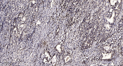
| Dilution | WB 1:500-2000;IHC-p 1:50-300 |
| Purification | The antibody was affinity-purified from rabbit serum by affinity-chromatography using specific immunogen. |
| Concentration | 1 mg/ml |
| Storage stability | -20°C/1 year |
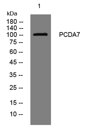
| Molecular Weight (Da) | 103kD |
| Observed band (KD) | |
| Background | This gene is a member of the protocadherin alpha gene cluster, one of three related gene clusters tandemly linked on chromosome five that demonstrate an unusual genomic organization similar to that of B-cell and T-cell receptor gene clusters. The alpha gene cluster is composed of 15 cadherin superfamily genes related to the mouse CNR genes and consists of 13 highly similar and 2 more distantly related coding sequences. The tandem array of 15 N-terminal exons, or variable exons, are followed by downstream C-terminal exons, or constant exons, which are shared by all genes in the cluster. The large, uninterrupted N-terminal exons each encode six cadherin ectodomains while the C-terminal exons encode the cytoplasmic domain. These neural cadherin-like cell adhesion proteins are integral plasma membrane proteins that most likely play a critical role in the establishment and function of specific cell-cell connections in the brain. Alternative splicing has been observed and additional variants have been suggested but their full-length nature has yet to be determined. [provided by RefSeq, Jul 2008], |
| Function | function:Potential calcium-dependent cell-adhesion protein. May be involved in the establishment and maintenance of specific neuronal connections in the brain.,similarity:Contains 6 cadherin domains., |
| Subcellular location | Cell membrane ; Single-pass type I membrane protein . |
| Expression |

Use collapsible tabs for more detailed information that will help customers make a purchasing decision.
Ex: Shipping and return policies, size guides, and other common questions.