 Français
 English
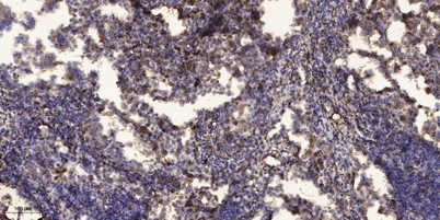
Rad17 (phospho Ser645) rabbit pAb
AO-06-ES1425-100

| ELK.No | ES1425 |
| Product name | Rad17 (phospho Ser645) rabbit pAb |
| Reactivity | Human;Mouse |
| Applications | WB; ELISA;IHC |
| Other name | RAD17; R24L; Cell cycle checkpoint protein RAD17; hRad17; RF-C/activator 1 homolog |
| Size | 100μL |
| Unit price ($) | 248 |
| Human gene ID | 5884 |
| Human Swiss-Prot | O75943 |
| Source | Rabbit |
| Isotype | IgG |
| Target | Rad17 |
| Fields | |
| Gene name | RAD17 |
| Protein name | Cell cycle checkpoint protein RAD17 |
| Human gene link | View Human Gene |
| Human Swiss link | View Human Swiss-Prot |
| Mouse gene ID | 19356 |
| Mouse gene link | View Mouse Gene |
| Mouse Swiss-Prot | Q6NXW6 |
| Mouse Swiss link | View Mouse Swiss-Prot |
| Rat gene ID | |
| Rat gene link | |
| Rat Swiss-Prot | |
| Rat Swiss link | |
| Immunogen | The antiserum was produced against synthesized peptide derived from human RAD17 around the phosphorylation site of Ser645. AA range:621-670 |
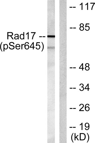
| Specificity | Phospho-Rad17 (S645) Polyclonal Antibody detects endogenous levels of Rad17 protein only when phosphorylated at S645. |
| Formulation | Liquid in PBS containing 50% glycerol, 0.5% BSA and 0.02% sodium azide. |
| Clonality | Polyclonal |
| Dilution | WB 1:500-2000;IHC-p 1:50-300; ELISA 2000-20000 |
| Purification | The antibody was affinity-purified from rabbit antiserum by affinity-chromatography using epitope-specific immunogen. |
| Concentration | 1 mg/ml |
| Storage stability | -20°C/1 year |
| Molecular Weight (Da) | |
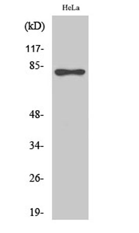
| Observed band (KD) | 77kD |
| Background | The protein encoded by this gene is highly similar to the gene product of Schizosaccharomyces pombe rad17, a cell cycle checkpoint gene required for cell cycle arrest and DNA damage repair in response to DNA damage. This protein shares strong similarity with DNA replication factor C (RFC), and can form a complex with RFCs. This protein binds to chromatin prior to DNA damage and is phosphorylated by the checkpoint kinase ATR following damage. This protein recruits the RAD1-RAD9-HUS1 checkpoint protein complex onto chromatin after DNA damage, which may be required for its phosphorylation. The phosphorylation of this protein is required for the DNA-damage-induced cell cycle G2 arrest, and is thought to be a critical early event during checkpoint signaling in DNA-damaged cells. Multiple alternatively spliced transcript variants of this gene, which encode four distinct protein isoforms, h |
| Function | function:Essential for sustained cell growth, maintenance of chromosomal stability, and ATR-dependent checkpoint activation upon DNA damage. Has a weak ATPase activity required for binding to chromatin. Participates in the recruitment of the RAD1-RAD9-HUS1 complex onto chromatin, and in CHEK1 activation. May also serve as a sensor of DNA replication progression, and may be involved in homologous recombination.,induction:By X-ray irradiation (isoform 1, isoform 3 and isoform 4).,PTM:Phosphorylated. Phosphorylation on Ser-646 and Ser-656 is cell cycle-regulated, enhanced by genotoxic stress, and required for activation of checkpoint signaling. Phosphorylation is mediated by ATR upon UV or replication arrest, whereas it may be mediated both by ATR and ATM upon ionizing radiation. Phosphorylation on both sites is required for interaction with RAD1 but dispensable for interaction with RFC3 or |
| Subcellular location | Nucleus . Phosphorylated form redistributes to discrete nuclear foci upon DNA damage. |
| Expression | Overexpressed in various cancer cell lines and in colon carcinoma (at protein level). Isoform 2 and isoform 3 are the most abundant isoforms in non irradiated cells (at protein level). Ubiquitous at low levels. Highly expressed in testis, where it is expressed within the germinal epithelium of the seminiferous tubuli. Weakly expressed in seminomas (testicular tumors). |

Use collapsible tabs for more detailed information that will help customers make a purchasing decision.
Ex: Shipping and return policies, size guides, and other common questions.