 Français
 English
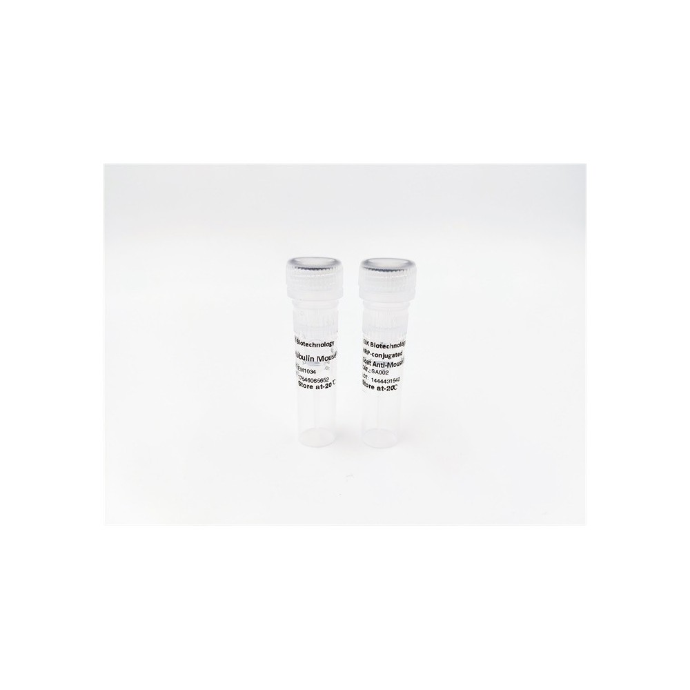
IGF-IR (phospho Tyr1165/Y1166) rabbit pAb
AO-06-ES1335-50

| ELK.No | ES1335 |
| Product name | IGF-IR (phospho Tyr1165/Y1166) rabbit pAb |
| Reactivity | Human;Mouse;Rat;Monkey |
| Applications | WB;ELISA |
| Other name | IGF1R; Insulin-like growth factor 1 receptor; Insulin-like growth factor I receptor; IGF-I receptor; CD antigen CD221 |
| Size | 50μL |
| Unit price ($) | 148 |
| Human gene ID | 3480 |
| Human Swiss-Prot | P08069 |
| Source | Rabbit |
| Isotype | IgG |
| Target | IGF-1R |
| Fields | >>EGFR tyrosine kinase inhibitor resistance;>>Endocrine resistance;>>MAPK signaling pathway;>>Ras signaling pathway;>>Rap1 signaling pathway;>>HIF-1 signaling pathway;>>FoxO signaling pathway;>>Oocyte meiosis;>>Autophagy - animal;>>Endocytosis;>>mTOR signaling pathway;>>PI3K-Akt signaling pathway;>>AMPK signaling pathway;>>Longevity regulating pathway;>>Longevity regulating pathway - multiple species;>>Focal adhesion;>>Adherens junction;>>Signaling pathways regulating pluripotency of stem cells;>>Long-term depression;>>Ovarian steroidogenesis;>>Progesterone-mediated oocyte maturation;>>Pathways in cancer;>>Transcriptional misregulation in cancer;>>Proteoglycans in cancer;>>Glioma;>>Prostate cancer;>>Melanoma;>>Breast cancer;>>Hepatocellular carcinoma |
| Gene name | IGF1R |
| Protein name | Insulin-like growth factor 1 receptor |
| Human gene link | View Human Gene |
| Human Swiss link | View Human Swiss-Prot |
| Mouse gene ID | 16001 |
| Mouse gene link | View Mouse Gene |
| Mouse Swiss-Prot | Q60751 |
| Mouse Swiss link | View Mouse Swiss-Prot |
| Rat gene ID | 25718 |
| Rat gene link | View Rat Gene |
| Rat Swiss-Prot | P24062 |
| Rat Swiss link | View Rat Swiss-Prot |
| Immunogen | The antiserum was produced against synthesized peptide derived from human IGF1R around the phosphorylation site of Tyr1165/Tyr1166. AA range:1131-1180 |
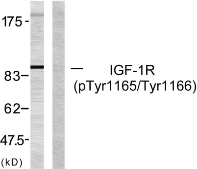
| Specificity | Phospho-IGF-IR (Y1165/Y1166) Polyclonal Antibody detects endogenous levels of IGF-IR protein only when phosphorylated at Y1165/Y1166. |
| Formulation | Liquid in PBS containing 50% glycerol, 0.5% BSA and 0.02% sodium azide. |
| Clonality | Polyclonal |
| Dilution | Western Blot: 1/500 - 1/2000. ELISA: 1/20000. Not yet tested in other applications. |
| Purification | The antibody was affinity-purified from rabbit antiserum by affinity-chromatography using epitope-specific immunogen. |
| Concentration | 1 mg/ml |
| Storage stability | -20°C/1 year |
| Molecular Weight (Da) | |
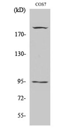
| Observed band (KD) | pro: 155kD, recetor beta: 95kD |
| Background | This receptor binds insulin-like growth factor with a high affinity. It has tyrosine kinase activity. The insulin-like growth factor I receptor plays a critical role in transformation events. Cleavage of the precursor generates alpha and beta subunits. It is highly overexpressed in most malignant tissues where it functions as an anti-apoptotic agent by enhancing cell survival. Alternatively spliced transcript variants encoding distinct isoforms have been found for this gene. [provided by RefSeq, May 2014], |
| Function | catalytic activity:ATP + a [protein]-L-tyrosine = ADP + a [protein]-L-tyrosine phosphate.,disease:Defects in IGF1R may be a cause in some cases of resistance to insulin-like growth factor 1 (IGF1 resistance) [MIM:270450]. IGF1 resistance is a gowth deficiency disorder characterized by intrauterine growth retardation and poor postnatal growth accompanied with increased plasma IGF1.,enzyme regulation:Autophosphorylation activates the kinase activity.,function:This receptor binds insulin-like growth factor 1 (IGF1) with a high affinity and IGF2 with a lower affinity. It has a tyrosine-protein kinase activity, which is necessary for the activation of the IGF1-stimulated downstream signaling cascade. When present in a hybrid receptor with INSR, binds IGF1. PubMed:12138094 shows that hybrid receptors composed of IGF1R and INSR isoform Long are activated with a high affinity by IGF1, with low a |
| Subcellular location | Cell membrane ; Single-pass type I membrane protein . |
| Expression | Found as a hybrid receptor with INSR in muscle, heart, kidney, adipose tissue, skeletal muscle, hepatoma, fibroblasts, spleen and placenta (at protein level). Expressed in a variety of tissues. Overexpressed in tumors, including melanomas, cancers of the colon, pancreas prostate and kidney. |


Use collapsible tabs for more detailed information that will help customers make a purchasing decision.
Ex: Shipping and return policies, size guides, and other common questions.
