 Français
 English

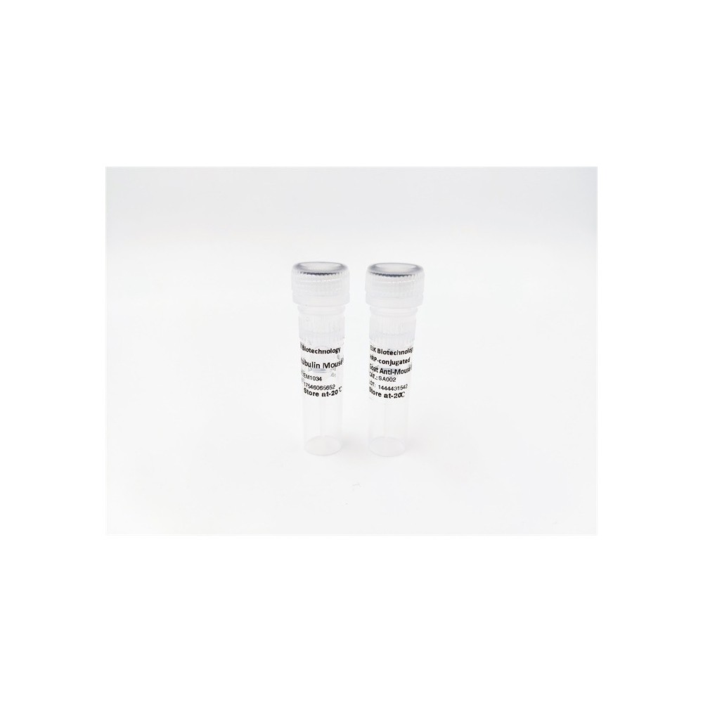

| ELK.No | ES14640 |
| Product name | MXRA8 rabbit pAb |
| Reactivity | Human; Mouse;Rat |
| Applications | WB;IHC |
| Other name | |
| Size | 50μL |
| Unit price ($) | 148 |
| Human gene ID | 54587 |
| Human Swiss-Prot | Q9BRK3 |
| Source | Rabbit |
| Isotype | IgG |
| Target | MXRA8 |
| Fields | |
| Gene name | MXRA8 |
| Protein name | MXRA8 |
| Human gene link | View Human Gene |
| Human Swiss link | View Human Swiss-Prot |
| Mouse gene ID | 74761 |
| Mouse gene link | View Mouse Gene |
| Mouse Swiss-Prot | Q9DBV4 |
| Mouse Swiss link | View Mouse Swiss-Prot |
| Rat gene ID | 313770 |
| Rat gene link | View Rat Gene |
| Rat Swiss-Prot | Q5XI43 |
| Rat Swiss link | View Rat Swiss-Prot |
| Immunogen | Synthesized peptide derived from human MXRA8 AA range: 42-92 |
| Specificity | This antibody detects endogenous levels of MXRA8 at Human/Mouse/Rat |
| Formulation | Liquid in PBS containing 50% glycerol, 0.5% BSA and 0.02% sodium azide. |
| Clonality | Polyclonal |
| Dilution | WB 1:500-2000;IHC-p 1:50-300 |
| Purification | The antibody was affinity-purified from rabbit serum by affinity-chromatography using specific immunogen. |
| Concentration | 1 mg/ml |
| Storage stability | -20°C/1 year |
| Molecular Weight (Da) | 49kD |
| Observed band (KD) | |
| Background | |
| Function | function:May play a role in the maturation and maintenance of blood-brain barrier.,similarity:Contains 2 Ig-like V-type (immunoglobulin-like) domains., |
| Subcellular location | Cell membrane ; Single-pass type I membrane protein . Cell junction, tight junction . Cytoplasm . Cell projection, cilium membrane . Nucleus . Primarily localizes to the cell membrane (PubMed:23386276). Detected in the cilium of primary chondrocytes (By similarity). Highly expressed at areas of cell-cell contact and may localize to tight junctions (By similarity). Also found in the nucleus where it is detected in the soluble (as opposed to chromatin-bound) fraction (PubMed:23386276). . |
| Expression | Detected in endothelial cells in mammary tissue, in both large vessels (left internal mammary artery) and small capillaries (vasa vasorum of the adventitia). |



Use collapsible tabs for more detailed information that will help customers make a purchasing decision.
Ex: Shipping and return policies, size guides, and other common questions.