 Français
 English

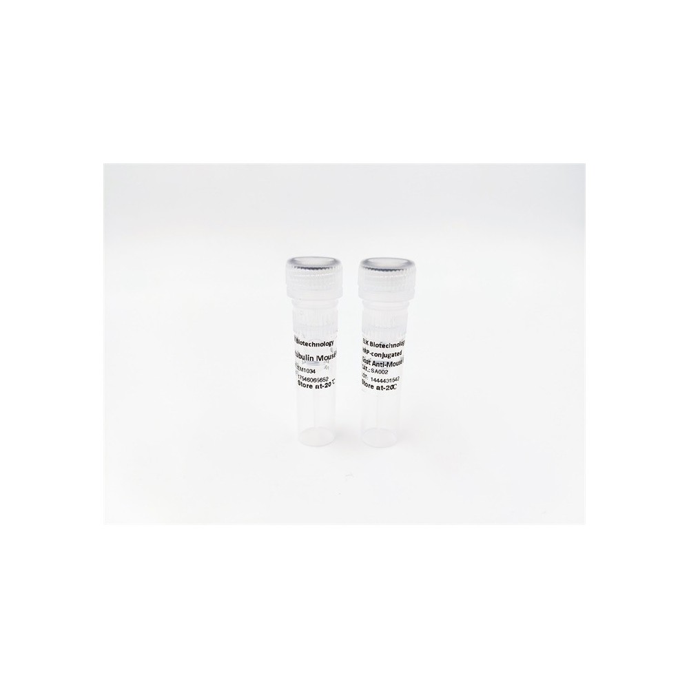

| ELK.No | ES15310 |
| Product name | KIF15 rabbit pAb |
| Reactivity | Human; Mouse;Rat |
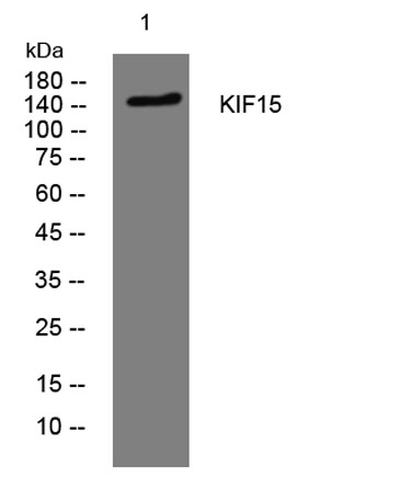
| Applications | WB |
| Other name | |
| Size | 100μL |
| Unit price ($) | 248 |
| Human gene ID | 56992 |
| Human Swiss-Prot | Q9NS87 |
| Source | Rabbit |
| Isotype | IgG |
| Target | KIF15 |
| Fields | |
| Gene name | KIF15 KLP2 KNSL7 |
| Protein name | KIF15 |
| Human gene link | View Human Gene |
| Human Swiss link | View Human Swiss-Prot |
| Mouse gene ID | 209737 |
| Mouse gene link | View Mouse Gene |
| Mouse Swiss-Prot | Q6P9L6 |
| Mouse Swiss link | View Mouse Swiss-Prot |
| Rat gene ID | 353302 |
| Rat gene link | View Rat Gene |
| Rat Swiss-Prot | Q7TSP2 |
| Rat Swiss link | View Rat Swiss-Prot |
| Immunogen | Synthesized peptide derived from human KIF15 AA range: 974-1024 |
| Specificity | This antibody detects endogenous levels of KIF15 at Human/Mouse/Rat |
| Formulation | Liquid in PBS containing 50% glycerol, 0.5% BSA and 0.02% sodium azide. |
| Clonality | Polyclonal |
| Dilution | WB 1:500-2000 |
| Purification | The antibody was affinity-purified from rabbit serum by affinity-chromatography using specific immunogen. |
| Concentration | 1 mg/ml |
| Storage stability | -20°C/1 year |
| Molecular Weight (Da) | 153kD |
| Observed band (KD) | |
| Background | |
| Function | function:Plus-end directed kinesin-like motor enzyme involved in mitotic spindle assembly.,similarity:Belongs to the kinesin-like protein family. KLP2 subfamily.,similarity:Contains 1 kinesin-motor domain.,subcellular location:Detected during the interphase in the cytoplasm as finely punctuate pattern and irregularly shaped dots. Detected during mitosis on the mitotic spindle. Colocalizes with TPX2 in mitosis. Localizes at the central spindle at anaphase (By similarity). Localizes at the sites of invaginating cell membranes, a position that corresponds to the location of the contractile actomyosin ring of dividing cells (By similarity). Colocalizes with actin in interphase (By similarity). Colocalizes in dendrites and in growth cone of axons with microtubules.,subunit:Interacts with MKI67 and TPX2.,tissue specificity:Expressed in testis, colon, tymus and in breast cancer., |
| Subcellular location | Cytoplasm. Cytoplasm, cytoskeleton, spindle. Detected during the interphase in the cytoplasm as finely punctuate pattern and irregularly shaped dots. Detected during mitosis on the mitotic spindle. Colocalizes with TPX2 in mitosis. Localizes at the central spindle at anaphase (By similarity). Localizes at the sites of invaginating cell membranes, a position that corresponds to the location of the contractile actomyosin ring of dividing cells (By similarity). Colocalizes with actin in interphase (By similarity). Colocalizes in dendrites and in growth cone of axons with microtubules (By similarity). . |
| Expression | Expressed in testis, colon, thymus and in breast cancer. |

Use collapsible tabs for more detailed information that will help customers make a purchasing decision.
Ex: Shipping and return policies, size guides, and other common questions.