 Français
 English


| ELK.No | ES15809 |
| Product name | HCP5 rabbit pAb |
| Reactivity | Human;Rat;Mouse; |
| Applications | WB |
| Other name | |
| Size | 100μL |
| Unit price ($) | 248 |
| Human gene ID | |
| Human Swiss-Prot | Q6MZN7 |
| Source | Rabbit |
| Isotype | IgG |
| Target | HCP5 |
| Fields | |
| Gene name | HCP5 |
| Protein name | HCP5 |
| Human gene link | |
| Human Swiss link | View Human Swiss-Prot |
| Mouse gene ID | |
| Mouse gene link | |
| Mouse Swiss-Prot | |
| Mouse Swiss link | |
| Rat gene ID | |
| Rat gene link | |
| Rat Swiss-Prot | |
| Rat Swiss link | |
| Immunogen | Synthesized peptide derived from human HCP5 AA range: 82-132 |
| Specificity | This antibody detects endogenous levels of HCP5 at Human |
| Formulation | Liquid in PBS containing 50% glycerol, 0.5% BSA and 0.02% sodium azide. |
| Clonality | Polyclonal |
| Dilution | WB 1:500-2000 |
| Purification | The antibody was affinity-purified from rabbit serum by affinity-chromatography using specific immunogen. |
| Concentration | 1 mg/ml |
| Storage stability | -20°C/1 year |
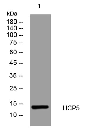
| Molecular Weight (Da) | 15kD |
| Observed band (KD) | |
| Background | |
| Function | miscellaneous:HCP5 is localized within the MHC class I region, but is not structurally related to MHC class I genes. HCP5 is related in sequence to human endogenous retroviruses HERV-L and HERV-16. It has sequence homology with retroviral Pol genes in HERV retroviral element; thus, it is itself a good candidate to interact with HIV-1, possibly through an antisense mechanism against retroviral transcript.,miscellaneous:Variation Gly-112 is associated with low viral loads in untreated HIV patients. The level of circulating virus in the plasma of HIV patients (viral set point) varies among individuals during the nonsymptomatic phase preceding the progression to AIDS. This polymorphism explains 9.6% of the total variation in set point and is associated with the HLA-B*5701 allele, which has the strongest described protective impact on HIV disease progression. However, it is possible that HPC5 |
| Subcellular location | |
| Expression | Expressed in lymphoid tissues; Detected in spleen as well as in B-cell lines, NK cell lines and activated lymphocytes. |

Use collapsible tabs for more detailed information that will help customers make a purchasing decision.
Ex: Shipping and return policies, size guides, and other common questions.