 Français
 English

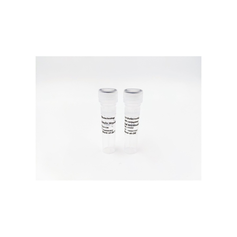

| ELK.No | ES15854 |
| Product name | GUC1A rabbit pAb |
| Reactivity | Human; Mouse |
| Applications | WB |
| Other name | |
| Size | 50μL |
| Unit price ($) | 148 |
| Human gene ID | 2978 |
| Human Swiss-Prot | P43080 |
| Source | Rabbit |
| Isotype | IgG |
| Target | GUC1A |
| Fields | >>Phototransduction |
| Gene name | GUCA1A C6orf131 GCAP GCAP1 GUCA1 |
| Protein name | GUC1A |
| Human gene link | View Human Gene |
| Human Swiss link | View Human Swiss-Prot |
| Mouse gene ID | 14913 |
| Mouse gene link | View Mouse Gene |
| Mouse Swiss-Prot | P43081 |
| Mouse Swiss link | View Mouse Swiss-Prot |
| Rat gene ID | |
| Rat gene link | |
| Rat Swiss-Prot | |
| Rat Swiss link | |
| Immunogen | Synthesized peptide derived from human GUC1A AA range: 63-113 |
| Specificity | This antibody detects endogenous levels of GUC1A at Human/Mouse |
| Formulation | Liquid in PBS containing 50% glycerol, 0.5% BSA and 0.02% sodium azide. |
| Clonality | Polyclonal |
| Dilution | WB 1:500-2000 |
| Purification | The antibody was affinity-purified from rabbit serum by affinity-chromatography using specific immunogen. |
| Concentration | 1 mg/ml |
| Storage stability | -20°C/1 year |
| Molecular Weight (Da) | 22kD |
| Observed band (KD) | |
| Background | This gene encodes an enzyme that plays a role in the recovery of retinal photoreceptors from photobleaching. This enzyme promotes the activity of retinal guanylyl cyclase-1 (GC1) at low calcium concentrations and inhibits GC1 at high calcium concentrations. Mutations in this gene can cause cone dystrophy 3 and code-rod dystrophy 14. Alternative splicing results in multiple transcript variants. [provided by RefSeq, Jan 2016], |
| Function | disease:Defects in GUCA1A are the cause of cone dystrophy type 3 (COD3) [MIM:602093]. COD3 is an autosomal dominant cone dystrophy. Cone dystrophies are retinal dystrophies characterized by progressive degeneration of the cone photoreceptors with preservation of rod function, as indicated by electroretinogram. However, some rod involvement may be present in some cone dystrophies, particularly at late stage. Affected individuals suffer from photophobia, loss of visual acuity, color vision and central visual field. Another sign is the absence of macular lesions for many years. Cone dystrophies are distinguished from the cone-rod dystrophies, in which some loss of peripheral vision also occurs.,function:Stimulates guanylyl cyclase 1 (GC1) when free calcium ions concentration is low and inhibits GC1 when free calcium ions concentration is elevated. This Ca(2+)-sensitive regulation of GC is a |
| Subcellular location | Membrane; Lipid-anchor . Photoreceptor inner segment . Cell projection, cilium, photoreceptor outer segment . Present at higher levels in cone than in rod outer segments (PubMed:9620085). Subcellular location is not affected by light or dark conditions. . |
| Expression | In the retina, it is expressed in rod and cone photoreceptors. |


Use collapsible tabs for more detailed information that will help customers make a purchasing decision.
Ex: Shipping and return policies, size guides, and other common questions.