 Français
 English

| ELK.No | ES1622 |
| Product name | AKAP 79 rabbit pAb |
| Reactivity | Human;Rat;Mouse; |
| Applications | WB;IHC;IF;ELISA |
| Other name | AKAP5; AKAP79; A-kinase anchor protein 5; AKAP-5; A-kinase anchor protein 79 kDa; AKAP 79; H21; cAMP-dependent protein kinase regulatory subunit II high affinity-binding protein |
| Size | 100μL |
| Unit price ($) | 248 |
| Human gene ID | 9495 |
| Human Swiss-Prot | P24588 |
| Source | Rabbit |
| Isotype | IgG |
| Target | AKAP 79 |
| Fields | |
| Gene name | AKAP5 |
| Protein name | A-kinase anchor protein 5 |
| Human gene link | View Human Gene |
| Human Swiss link | View Human Swiss-Prot |
| Mouse gene ID | |
| Mouse gene link | |
| Mouse Swiss-Prot | D3YVF0 |
| Mouse Swiss link | View Mouse Swiss-Prot |
| Rat gene ID | |
| Rat gene link | |
| Rat Swiss-Prot | |
| Rat Swiss link | |
| Immunogen | The antiserum was produced against synthesized peptide derived from human AKAP5. AA range:1-50 |
| Specificity | AKAP 79 Polyclonal Antibody detects endogenous levels of AKAP 79 protein. |
| Formulation | Liquid in PBS containing 50% glycerol, 0.5% BSA and 0.02% sodium azide. |
| Clonality | Polyclonal |
| Dilution | Western Blot: 1/500 - 1/2000. Immunohistochemistry: 1/100 - 1/300. Immunofluorescence: 1/200 - 1/1000. ELISA: 1/10000. Not yet tested in other applications. |
| Purification | The antibody was affinity-purified from rabbit antiserum by affinity-chromatography using epitope-specific immunogen. |
| Concentration | 1 mg/ml |
| Storage stability | -20°C/1 year |
| Molecular Weight (Da) | |
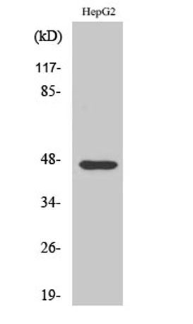
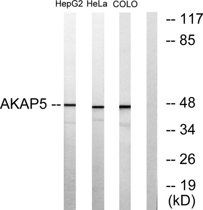
| Observed band (KD) | 47kD |
| Background | The A-kinase anchor proteins (AKAPs) are a group of structurally diverse proteins, which have the common function of binding to the regulatory subunit of protein kinase A (PKA) and confining the holoenzyme to discrete locations within the cell. This gene encodes a member of the AKAP family. The encoded protein binds to the RII-beta regulatory subunit of PKA, and also to protein kinase C and the phosphatase calcineurin. It is predominantly expressed in cerebral cortex and may anchor the PKA protein at postsynaptic densities (PSD) and be involved in the regulation of postsynaptic events. It is also expressed in T lymphocytes and may function to inhibit interleukin-2 transcription by disrupting calcineurin-dependent dephosphorylation of NFAT. [provided by RefSeq, Jul 2008], |
| Function | domain:RII-alpha binding site, predicted to form an amphipathic helix, could participate in protein-protein interactions with a complementary surface on the R-subunit dimer.,function:May anchor the PKA protein to cytoskeletal and/or organelle-associated proteins, targeting the signal carried by cAMP to specific intracellular effectors. Association with to the beta2-adrenergic receptor (beta2-AR) not only regulates beta2-AR signaling pathway, but also the activation by PKA by switching off the beta2-AR signaling cascade.,miscellaneous:The N-terminal region, which is highly basic, is required for interaction with calmodulin.,similarity:Contains 1 AKAP domain.,subcellular location:Associated with particulate fractions.,subunit:Binding protein for dimer of the RII-beta regulatory subunit of cAMP-dependent protein kinase (PKA) and also for the protein kinase C (PKC) and the phosphatase calcin |
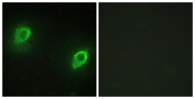
| Subcellular location | Postsynaptic recycling endosome membrane ; Lipid-anchor . Associates with lipid rafts. . |
| Expression | Predominantly in the cerebral cortex and the postsynaptic densities of the forebrain, and to a lesser extent in adrenal medulla, lung and anterior pituitary. |

Use collapsible tabs for more detailed information that will help customers make a purchasing decision.
Ex: Shipping and return policies, size guides, and other common questions.