 Français
 English

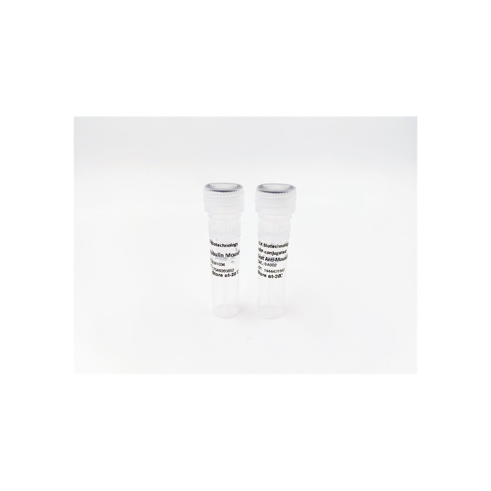

| ELK.No | ES16354 |
| Product name | FNDC1 rabbit pAb |
| Reactivity | Human;Rat |
| Applications | WB |
| Other name | |
| Size | 50μL |
| Unit price ($) | 148 |
| Human gene ID | 84624 |
| Human Swiss-Prot | Q4ZHG4 |
| Source | Rabbit |
| Isotype | IgG |
| Target | FNDC1 |
| Fields | |
| Gene name | FNDC1 FNDC2 KIAA1866 MEL4B3 |
| Protein name | FNDC1 |
| Human gene link | View Human Gene |
| Human Swiss link | View Human Swiss-Prot |
| Mouse gene ID | |
| Mouse gene link | |
| Mouse Swiss-Prot | |
| Mouse Swiss link | |
| Rat gene ID | 308099 |
| Rat gene link | View Rat Gene |
| Rat Swiss-Prot | Q2Q0I9 |
| Rat Swiss link | View Rat Swiss-Prot |
| Immunogen | Synthesized peptide derived from human FNDC1 AA range: 988-1038 |
| Specificity | This antibody detects endogenous levels of FNDC1 at Human/Rat |
| Formulation | Liquid in PBS containing 50% glycerol, 0.5% BSA and 0.02% sodium azide. |
| Clonality | Polyclonal |
| Dilution | WB 1:500-2000 |
| Purification | The antibody was affinity-purified from rabbit serum by affinity-chromatography using specific immunogen. |
| Concentration | 1 mg/ml |
| Storage stability | -20°C/1 year |
| Molecular Weight (Da) | 208kD |
| Observed band (KD) | |
| Background | |
| Function | caution:It is uncertain whether Met-1 or Met-53 is the initiator.,function:May be an activator of G protein signaling.,induction:By TGF-beta present in the melanoma cell conditioned medium (MCCM).,similarity:Contains 5 fibronectin type-III domains.,tissue specificity:Almost absent from healthy skin; especially in epidermal keratinocytes, skin fibroblasts or endothelial cells and is barely detectable in benign melanocytic naevi. Expressed in the stroma close to skin tumors, in the tumor cells themselves and in the epidermis of psoriasis., |
| Subcellular location | Secreted . |
| Expression | Almost absent from healthy skin; especially in epidermal keratinocytes, skin fibroblasts or endothelial cells and is barely detectable in benign melanocytic naevi. Expressed in the stroma close to skin tumors, in the tumor cells themselves and in the epidermis of psoriasis. |


Use collapsible tabs for more detailed information that will help customers make a purchasing decision.
Ex: Shipping and return policies, size guides, and other common questions.