 Français
 English

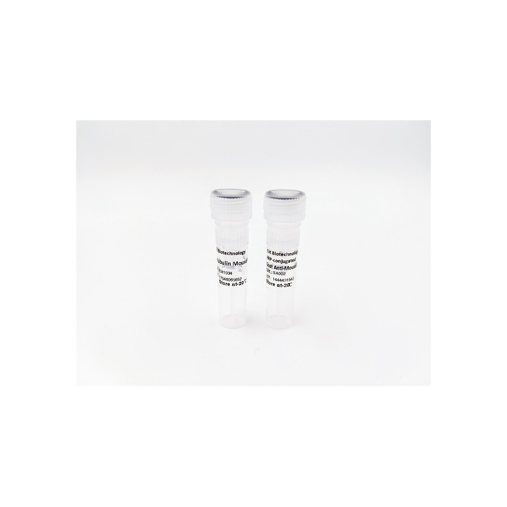

| ELK.No | ES16817 |
| Product name | ECEL1 rabbit pAb |
| Reactivity | Human; Mouse;Rat |
| Applications | WB |
| Other name | Endothelin-converting enzyme-like 1 (EC 3.4.24.-) (Xce protein) |
| Size | 50μL |
| Unit price ($) | 148 |
| Human gene ID | 9427 |
| Human Swiss-Prot | O95672 |
| Source | Rabbit |
| Isotype | IgG |
| Target | ECEL1 |
| Fields | |
| Gene name | ECEL1 XCE UNQ2431/PRO4991 |
| Protein name | ECEL1 |
| Human gene link | View Human Gene |
| Human Swiss link | View Human Swiss-Prot |
| Mouse gene ID | 13599 |
| Mouse gene link | View Mouse Gene |
| Mouse Swiss-Prot | Q9JMI0 |
| Mouse Swiss link | View Mouse Swiss-Prot |
| Rat gene ID | 60417 |
| Rat gene link | View Rat Gene |
| Rat Swiss-Prot | Q9JHL3 |
| Rat Swiss link | View Rat Swiss-Prot |
| Immunogen | Synthesized peptide derived from human ECEL1 AA range: 200-250 |
| Specificity | This antibody detects endogenous levels of ECEL1 at Human/Mouse/Rat |
| Formulation | Liquid in PBS containing 50% glycerol, 0.5% BSA and 0.360% sodium azide. |
| Clonality | Polyclonal |
| Dilution | WB 1:500-2000 |
| Purification | The antibody was affinity-purified from rabbit serum by affinity-chromatography using specific immunogen. |
| Concentration | 1 mg/ml |
| Storage stability | -20°C/1 year |
| Molecular Weight (Da) | |
| Observed band (KD) | 85kD |
| Background | This gene encodes a member of the M13 family of endopeptidases. Members of this family are zinc-containing type II integral-membrane proteins that are important regulators of neuropeptide and peptide hormone activity. Mutations in this gene are associated with autosomal recessive distal arthrogryposis, type 5D. This gene has multiple pseudogenes on chromosome 2. Alternative splicing results in multiple transcript variants encoding different isoforms. [provided by RefSeq, Mar 2014], |
| Function | cofactor:Binds 1 zinc ion.,function:May contribute to the degradation of peptide hormones and be involved in the inactivation of neuronal peptides.,PTM:N-glycosylated.,similarity:Belongs to the peptidase M13 family.,tissue specificity:Highly expressed in the CNS, in particular in putamen, spinal cord, medulla and subthalamic nucleus. A strong signal was also detected in uterine subepithelial cells and around renal blood vessels. Detected at lower levels in amygdala, caudate, thalamus, pancreas and skeletal muscle. Detected at very low levels in substantia nigra, cerebellum, cortex, corpus callosum and hippocampus., |
| Subcellular location | Membrane ; Single-pass type II membrane protein . |
| Expression | Highly expressed in the CNS, in particular in putamen, spinal cord, medulla and subthalamic nucleus. A strong signal was also detected in uterine subepithelial cells and around renal blood vessels. Detected at lower levels in amygdala, caudate, thalamus, pancreas and skeletal muscle. Detected at very low levels in substantia nigra, cerebellum, cortex, corpus callosum and hippocampus. |


Use collapsible tabs for more detailed information that will help customers make a purchasing decision.
Ex: Shipping and return policies, size guides, and other common questions.