 Français
 English

| ELK.No | ES1701 |
| Product name | ARK-2 rabbit pAb |
| Reactivity | Human;Mouse;Rat;Monkey |
| Applications | WB;IHC;IF;ELISA |
| Other name | AURKB; AIK2; AIM1; AIRK2; ARK2; STK1; STK12; STK5; Aurora kinase B; Aurora 1; Aurora- and IPL1-like midbody-associated protein 1; AIM-1; Aurora/IPL1-related kinase 2; ARK-2; Aurora-related kinase 2; STK-1; Serine/threonine-protein kinase 12 |
| Size | 50μL |
| Unit price ($) | 148 |
| Human gene ID | 9212 |
| Human Swiss-Prot | Q96GD4 |
| Source | Rabbit |
| Isotype | IgG |
| Target | ARK-2 |
| Fields | |
| Gene name | AURKB |
| Protein name | Aurora kinase B |
| Human gene link | View Human Gene |
| Human Swiss link | View Human Swiss-Prot |
| Mouse gene ID | 20877 |
| Mouse gene link | View Mouse Gene |
| Mouse Swiss-Prot | O70126 |
| Mouse Swiss link | View Mouse Swiss-Prot |
| Rat gene ID | 114592 |
| Rat gene link | View Rat Gene |
| Rat Swiss-Prot | O55099 |
| Rat Swiss link | View Rat Swiss-Prot |
| Immunogen | The antiserum was produced against synthesized peptide derived from human AurB. AA range:198-247 |
| Specificity | ARK-2 Polyclonal Antibody detects endogenous levels of ARK-2 protein. |
| Formulation | Liquid in PBS containing 50% glycerol, 0.5% BSA and 0.02% sodium azide. |
| Clonality | Polyclonal |
| Dilution | Western Blot: 1/500 - 1/2000. Immunohistochemistry: 1/100 - 1/300. Immunofluorescence: 1/200 - 1/1000. ELISA: 1/10000. Not yet tested in other applications. |
| Purification | The antibody was affinity-purified from rabbit antiserum by affinity-chromatography using epitope-specific immunogen. |
| Concentration | 1 mg/ml |
| Storage stability | -20°C/1 year |
| Molecular Weight (Da) | |
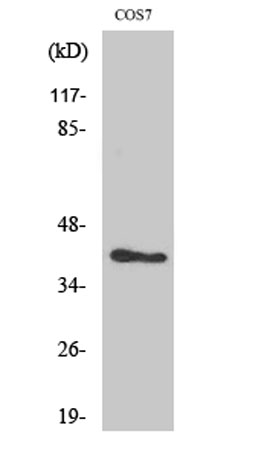
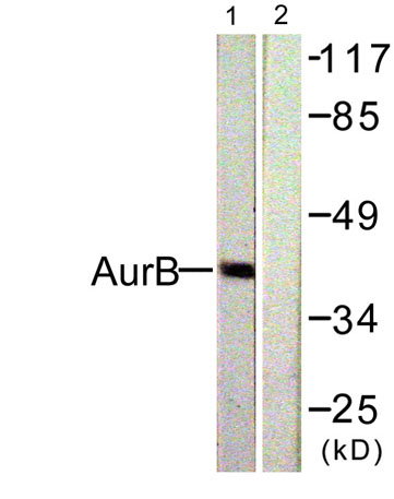
| Observed band (KD) | 39kD |
| Background | This gene encodes a member of the aurora kinase subfamily of serine/threonine kinases. The genes encoding the other two members of this subfamily are located on chromosomes 19 and 20. These kinases participate in the regulation of alignment and segregation of chromosomes during mitosis and meiosis through association with microtubules. A pseudogene of this gene is located on chromosome 8. Alternatively spliced transcript variants have been found for this gene. [provided by RefSeq, Sep 2015], |
| Function | catalytic activity:ATP + a protein = ADP + a phosphoprotein.,cofactor:Magnesium.,disease:Disruptive regulation of expression is a possibile mechanism of the perturbation of chromosomal integrity in cancer cells through its dominant-negative effect on cytokinesis.,function:May be directly involved in regulating the cleavage of polar spindle microtubules and is a key regulator for the onset of cytokinesis during mitosis. Component of the chromosomal passenger complex (CPC), a complex that acts as a key regulator of mitosis. The CPC complex has essential functions at the centromere in ensuring correct chromosome alignment and segregation and is required for chromatin-induced microtubule stabilization and spindle assembly. Phosphorylates 'Ser-10' and 'Ser-28' of histone H3 during mitosis.,similarity:Belongs to the protein kinase superfamily. Ser/Thr protein kinase family. Aurora subfamily.,s |
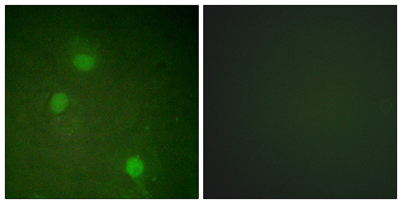
| Subcellular location | Nucleus . Chromosome . Chromosome, centromere . Chromosome, centromere, kinetochore . Cytoplasm, cytoskeleton, spindle . Midbody . Localizes on chromosome arms and inner centromeres from prophase through metaphase and then transferring to the spindle midzone and midbody from anaphase through cytokinesis (PubMed:20929775). Colocalized with gamma tubulin in the midbody (PubMed:17726514). Proper localization of the active, Thr-232-phosphorylated form during metaphase may be dependent upon interaction with SPDYC (PubMed:20605920). Colocalized with SIRT2 during cytokinesis with the midbody (PubMed:17726514). Localization (and probably targeting of the CPC) to the inner centromere occurs predominantly in regions with overlapping mitosis-specific histone phosphorylations H3pT3 and H2ApT12 (PubMed |
| Expression | High level expression seen in the thymus. It is also expressed in the spleen, lung, testis, colon, placenta and fetal liver. Expressed during S and G2/M phase and expression is up-regulated in cancer cells during M phase. ; [Isoform 3]: Not expressed in normal liver, high expression in metastatic liver. |

Use collapsible tabs for more detailed information that will help customers make a purchasing decision.
Ex: Shipping and return policies, size guides, and other common questions.