 Français
 English

| ELK.No | ES17345 |
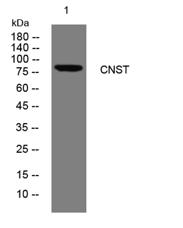
| Product name | CNST rabbit pAb |
| Reactivity | Human; Mouse |
| Applications | WB |
| Other name | |
| Size | 100μL |
| Unit price ($) | 248 |
| Human gene ID | 163882 |
| Human Swiss-Prot | Q6PJW8 |
| Source | Rabbit |
| Isotype | IgG |
| Target | CNST |
| Fields | |
| Gene name | CNST C1orf71 |
| Protein name | CNST |
| Human gene link | View Human Gene |
| Human Swiss link | View Human Swiss-Prot |
| Mouse gene ID | 226744 |
| Mouse gene link | View Mouse Gene |
| Mouse Swiss-Prot | Q8CBC4 |
| Mouse Swiss link | View Mouse Swiss-Prot |
| Rat gene ID | |
| Rat gene link | |
| Rat Swiss-Prot | |
| Rat Swiss link | |
| Immunogen | Synthesized peptide derived from human CNST AA range: 296-346 |
| Specificity | This antibody detects endogenous levels of CNST at Human/Mouse |
| Formulation | Liquid in PBS containing 50% glycerol, 0.5% BSA and 0.02% sodium azide. |
| Clonality | Polyclonal |
| Dilution | WB 1:500-2000 |
| Purification | The antibody was affinity-purified from rabbit serum by affinity-chromatography using specific immunogen. |
| Concentration | 1 mg/ml |
| Storage stability | -20°C/1 year |
| Molecular Weight (Da) | 80kD |
| Observed band (KD) | |
| Background | Targeting of numerous transmembrane proteins to the cell surface is thought to depend on their recognition by cargo receptors that interact with the adaptor machinery for anterograde traffic at the distal end of the Golgi complex. Consortin (CNST) is an integral membrane protein that acts as a binding partner of connexins, the building blocks of gap junctions, and acts as a trans-Golgi network (TGN) receptor involved in connexin targeting to the plasma membrane and recycling from the cell surface (del Castillo et al., 2010 [PubMed 19864490]).[supplied by OMIM, Jun 2010], |
| Function | |
| Subcellular location | Cell membrane ; Single-pass membrane protein . Golgi apparatus, trans-Golgi network membrane ; Single-pass membrane protein . Cytoplasmic vesicle, secretory vesicle . Located predominantly in the trans-Golgi network. Probably trafficks between the trans-Golgi network and the cell membrane via the secretory pathway. |
| Expression |

Use collapsible tabs for more detailed information that will help customers make a purchasing decision.
Ex: Shipping and return policies, size guides, and other common questions.