 Français
 English
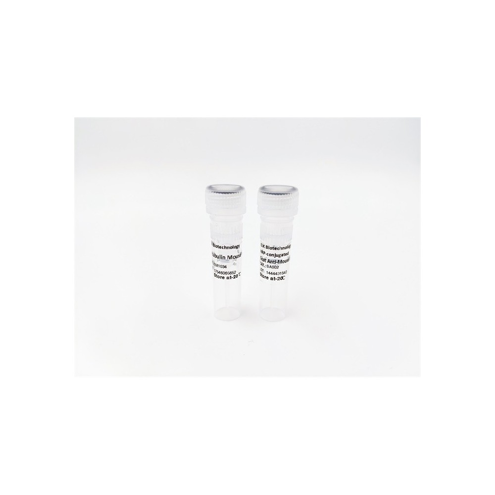

| ELK.No | ES17547 |
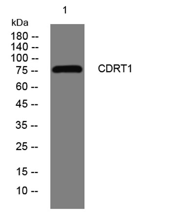
| Product name | CDRT1 rabbit pAb |
| Reactivity | Human;Rat;Mouse; |
| Applications | WB |
| Other name | |
| Size | 100μL |
| Unit price ($) | 248 |
| Human gene ID | 374286 |
| Human Swiss-Prot | O95170 |
| Source | Rabbit |
| Isotype | IgG |
| Target | CDRT1 |
| Fields | |
| Gene name | CDRT1 C17orf1 C17orf1A HREP |
| Protein name | CDRT1 |
| Human gene link | View Human Gene |
| Human Swiss link | View Human Swiss-Prot |
| Mouse gene ID | |
| Mouse gene link | |
| Mouse Swiss-Prot | |
| Mouse Swiss link | |
| Rat gene ID | |
| Rat gene link | |
| Rat Swiss-Prot | |
| Rat Swiss link | |
| Immunogen | Synthesized peptide derived from human CDRT1 AA range: 364-414 |
| Specificity | This antibody detects endogenous levels of CDRT1 at Human |
| Formulation | Liquid in PBS containing 50% glycerol, 0.5% BSA and 0.02% sodium azide. |
| Clonality | Polyclonal |
| Dilution | WB 1:500-2000 |
| Purification | The antibody was affinity-purified from rabbit serum by affinity-chromatography using specific immunogen. |
| Concentration | 1 mg/ml |
| Storage stability | -20°C/1 year |
| Molecular Weight (Da) | 83kD |
| Observed band (KD) | |
| Background | Members of the F-box protein family, such as FBXW10, are characterized by an approximately 40-amino acid F-box motif. SCF complexes, formed by SKP1 (MIM 601434), cullin (see CUL1; MIM 603034), and F-box proteins, act as protein-ubiquitin ligases. F-box proteins interact with SKP1 through the F box, and they interact with ubiquitination targets through other protein interaction domains (Jin et al., 2004 [PubMed 15520277]).[supplied by OMIM, Mar 2008], |
| Function | miscellaneous:CDRT1 gene is located centromeric to and partially within proximal CMT1A-REP element. CMT1A-REP is a complex binary repeat element flanking a 1.5-Mb DNA region duplicated in Charcot-Marie-Tooth disease type I (CMT1A) or deleted in Hereditary neuropathy (HNPP).,similarity:Contains 4 WD repeats.,tissue specificity:Expressed in pancreas, heart and skeletal muscle., |
| Subcellular location | intracellular, |
| Expression | Expressed in pancreas, heart and skeletal muscle. |

Use collapsible tabs for more detailed information that will help customers make a purchasing decision.
Ex: Shipping and return policies, size guides, and other common questions.