 Français
 English
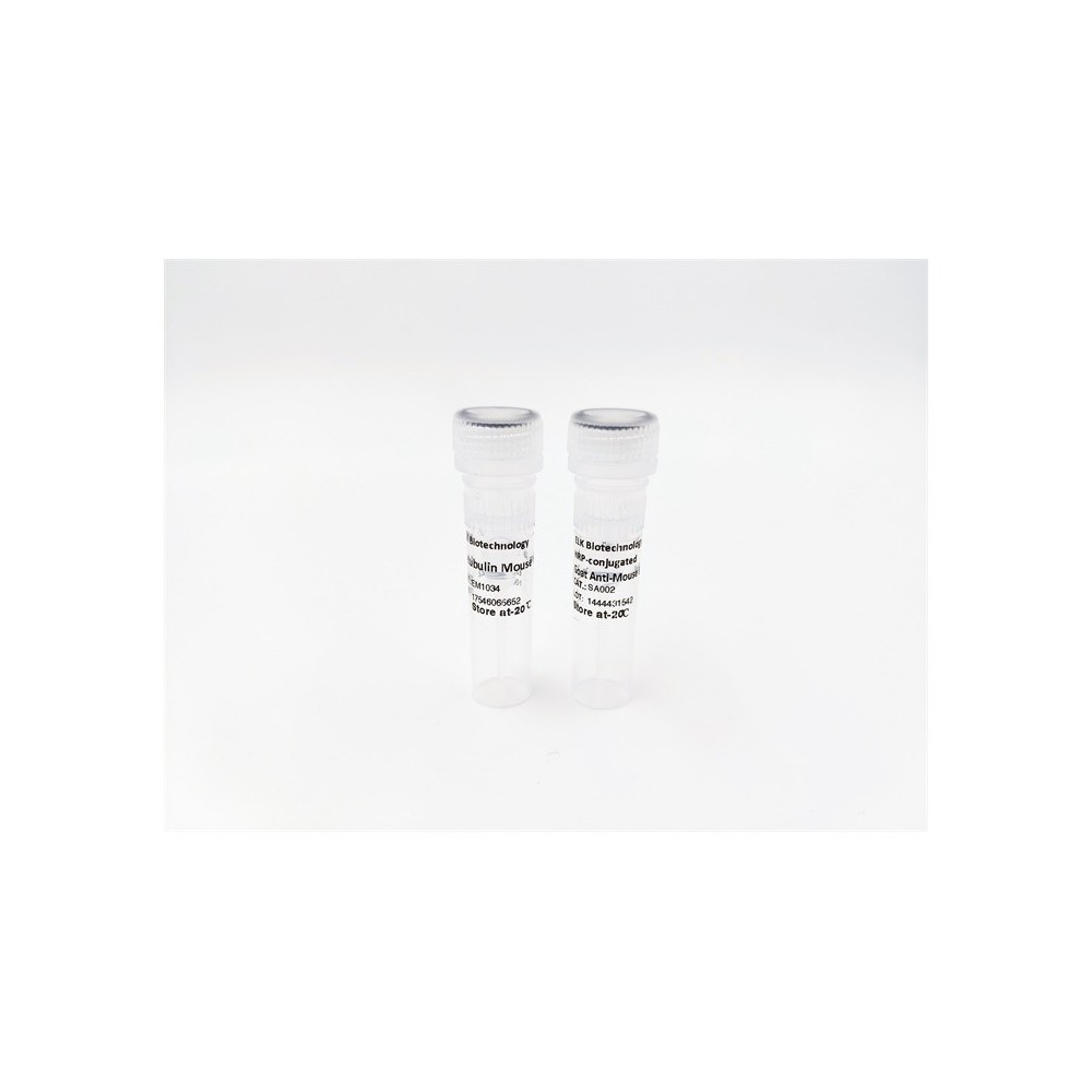

| ELK.No | ES1760 |
| Product name | Bcl-w rabbit pAb |
| Reactivity | Human;Mouse;Rat |
| Applications | WB;IHC;IF;ELISA |
| Other name | BCL2L2; BCLW; KIAA0271; Bcl-2-like protein 2; Bcl2-L-2; Apoptosis regulator Bcl-W |
| Size | 100μL |
| Unit price ($) | 248 |
| Human gene ID | 599 |
| Human Swiss-Prot | Q92843 |
| Source | Rabbit |
| Isotype | IgG |
| Target | Bcl-w |
| Fields | >>MicroRNAs in cancer |
| Gene name | BCL2L2 |
| Protein name | Bcl-2-like protein 2 |
| Human gene link | View Human Gene |
| Human Swiss link | View Human Swiss-Prot |
| Mouse gene ID | 12050 |
| Mouse gene link | View Mouse Gene |
| Mouse Swiss-Prot | P70345 |
| Mouse Swiss link | View Mouse Swiss-Prot |
| Rat gene ID | |
| Rat gene link | |
| Rat Swiss-Prot | |
| Rat Swiss link | |
| Immunogen | The antiserum was produced against synthesized peptide derived from human BCLW. AA range:131-180 |
| Specificity | Bcl-w Polyclonal Antibody detects endogenous levels of Bcl-w protein. |
| Formulation | Liquid in PBS containing 50% glycerol, 0.5% BSA and 0.02% sodium azide. |
| Clonality | Polyclonal |
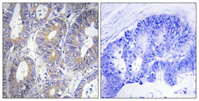
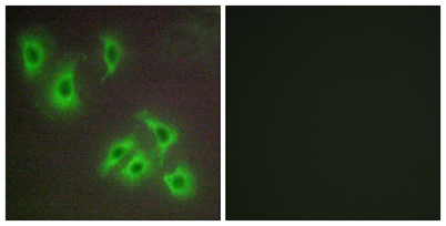
| Dilution | Western Blot: 1/500 - 1/2000. Immunohistochemistry: 1/100 - 1/300. Immunofluorescence: 1/200 - 1/1000. ELISA: 1/10000. Not yet tested in other applications. |
| Purification | The antibody was affinity-purified from rabbit antiserum by affinity-chromatography using epitope-specific immunogen. |
| Concentration | 1 mg/ml |
| Storage stability | -20°C/1 year |
| Molecular Weight (Da) | |
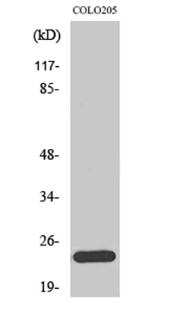
| Observed band (KD) | 25kD |
| Background | This gene encodes a member of the BCL-2 protein family. The proteins of this family form hetero- or homodimers and act as anti- and pro-apoptotic regulators. Expression of this gene in cells has been shown to contribute to reduced cell apoptosis under cytotoxic conditions. Studies of the related gene in mice indicated a role in the survival of NGF- and BDNF-dependent neurons. Mutation and knockout studies of the mouse gene demonstrated an essential role in adult spermatogenesis. Alternative splicing results in multiple transcript variants. Read-through transcription also exists between this gene and the neighboring downstream PABPN1 (poly(A) binding protein, nuclear 1) gene. [provided by RefSeq, Dec 2010], |
| Function | domain:The BH1 and BH2 motifs form a hydrophobic groove which acts as a docking site for the BH3 domain of some pro-apoptotic proteins. The C-terminal residues of BCL2L2 fold into the BH3-binding cleft and modulate pro-survival activity by regulating ligand access. When BH3 domain-containing proteins bind, they displace the C-terminus, allowing its insertion into the membrane and neutralizing the pro-survival activity of BCL2L2.,domain:The BH4 motif seems to be involved in the anti-apoptotic function.,function:Promotes cell survival. Blocks dexamethasone-induced apoptosis. Mediates survival of postmitotic Sertoli cells by suppressing death-promoting activity of BAX.,similarity:Belongs to the Bcl-2 family.,subcellular location:Loosely associated with the mitochondrial membrane in healthy cells. During apoptosis, tightly bound to the membrane.,tissue specificity:Expressed (at protein level |
| Subcellular location | Mitochondrion membrane ; Peripheral membrane protein . Loosely associated with the mitochondrial membrane in healthy cells. During apoptosis, tightly bound to the membrane. |
| Expression | Expressed (at protein level) in a wide range of tissues with highest levels in brain, spinal cord, testis, pancreas, heart, spleen and mammary glands. Moderate levels found in thymus, ovary and small intestine. Not detected in salivary gland, muscle or liver. Also expressed in cell lines of myeloid, fibroblast and epithelial origin. Not detected in most lymphoid cell lines. |

Use collapsible tabs for more detailed information that will help customers make a purchasing decision.
Ex: Shipping and return policies, size guides, and other common questions.