 Français
 English

| ELK.No | ES17927 |
| Product name | C99L2 rabbit pAb |
| Reactivity | Human; Mouse;Rat |
| Applications | WB |
| Other name | |
| Size | 50μL |
| Unit price ($) | 148 |
| Human gene ID | 83692 |
| Human Swiss-Prot | Q8TCZ2 |
| Source | Rabbit |
| Isotype | IgG |
| Target | C99L2 |
| Fields | >>Cell adhesion molecules;>>Leukocyte transendothelial migration |
| Gene name | CD99L2 MIC2L1 UNQ1964/PRO4486 |
| Protein name | C99L2 |
| Human gene link | View Human Gene |
| Human Swiss link | View Human Swiss-Prot |
| Mouse gene ID | 171486 |
| Mouse gene link | View Mouse Gene |
| Mouse Swiss-Prot | Q8BIF0 |
| Mouse Swiss link | View Mouse Swiss-Prot |
| Rat gene ID | 171485 |
| Rat gene link | View Rat Gene |
| Rat Swiss-Prot | Q8R1R5 |
| Rat Swiss link | View Rat Swiss-Prot |
| Immunogen | Synthesized peptide derived from human C99L2 AA range: 204-254 |
| Specificity | This antibody detects endogenous levels of C99L2 at Human/Mouse/Rat |
| Formulation | Liquid in PBS containing 50% glycerol, 0.5% BSA and 0.02% sodium azide. |
| Clonality | Polyclonal |
| Dilution | WB 1:500-2000 |
| Purification | The antibody was affinity-purified from rabbit serum by affinity-chromatography using specific immunogen. |
| Concentration | 1 mg/ml |
| Storage stability | -20°C/1 year |
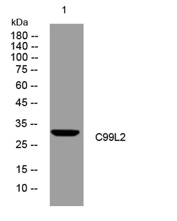
| Molecular Weight (Da) | 29kD |
| Observed band (KD) | |
| Background | This gene encodes a cell-surface protein that is similar to CD99. A similar protein in mouse functions as an adhesion molecule during leukocyte extravasation. Alternate splicing results in multiple transcript variants. [provided by RefSeq, May 2010], |
| Function | function:May function as a homophilic adhesion molecule. Functions in leukocyte-endothelial cell interactions during leukocyte extravasation, and in particular, at the diapedesis step. Does not seem to be involved in docking of leukocytes to the vessel wall or in lymphocyte diapedesis.,PTM:O-glycosylated.,similarity:Belongs to the CD99 family.,tissue specificity:Expressed in brain, heart, lung, liver, spleen, kidney, stomach, small intestine, skeletal muscle, ovary, thymus, testis and uterus. Lower expression seen in thymus. Expressed in E18 uterus and placenta., |
| Subcellular location | Cell membrane ; Single-pass type I membrane protein ; Extracellular side . Cell junction . |
| Expression | Expressed in many tissues, with low expression in thymus. |

Use collapsible tabs for more detailed information that will help customers make a purchasing decision.
Ex: Shipping and return policies, size guides, and other common questions.