 Français
 English
RhoA (Phospho Ser188) rabbit pAb
AO-06-ES20149-100

| ELK.No | ES20149 |
| Product name | RhoA (Phospho Ser188) rabbit pAb |
| Reactivity | Human;Mouse;Rat |
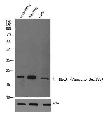
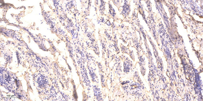
| Applications | WB;ELISA;IHC |
| Other name | Transforming protein RhoA (Rho cDNA clone 12;h12) |
| Size | 100μL |
| Unit price ($) | 248 |
| Human gene ID | 387 |
| Human Swiss-Prot | P61586 |
| Source | Rabbit |
| Isotype | IgG |
| Target | RhoA |
| Fields | >>Ras signaling pathway;>>Rap1 signaling pathway;>>cGMP-PKG signaling pathway;>>cAMP signaling pathway;>>Chemokine signaling pathway;>>Sphingolipid signaling pathway;>>Phospholipase D signaling pathway;>>Endocytosis;>>mTOR signaling pathway;>>Vascular smooth muscle contraction;>>Wnt signaling pathway;>>TGF-beta signaling pathway;>>Axon guidance;>>Focal adhesion;>>Adherens junction;>>Tight junction;>>Platelet activation;>>NOD-like receptor signaling pathway;>>C-type lectin receptor signaling pathway;>>T cell receptor signaling pathway;>>Leukocyte transendothelial migration;>>Neurotrophin signaling pathway;>>Regulation of actin cytoskeleton;>>Oxytocin signaling pathway;>>Parathyroid hormone synthesis, secretion and action;>>Pancreatic secretion;>>Bacterial invasion of epithelial cells;>>Pathogenic Escherichia coli infection;>>Shigellosis;>>Salmonella infection;>>Pertussis;>>Yersinia infection;>>Tuberculosis;>>Human cytomegalovirus infection;>>Pathways in cancer;>>Viral carcinogenesis;>>P |
| Gene name | RHOA ARH12 ARHA RHO12 |
| Protein name | RhoA (Phospho Ser188) |
| Human gene link | View Human Gene |
| Human Swiss link | View Human Swiss-Prot |
| Mouse gene ID | 11848 |
| Mouse gene link | View Mouse Gene |
| Mouse Swiss-Prot | Q9QUI0 |
| Mouse Swiss link | View Mouse Swiss-Prot |
| Rat gene ID | 117273 |
| Rat gene link | View Rat Gene |
| Rat Swiss-Prot | P61589 |
| Rat Swiss link | View Rat Swiss-Prot |
| Immunogen | Synthesized peptide derived from human RhoA (Phospho Ser188) |
| Specificity | This antibody detects endogenous levels of Human,Mouse,Rat RhoA (Phospho Ser188) |
| Formulation | Liquid in PBS containing 50% glycerol, 0.5% BSA and 0.02% sodium azide. |
| Clonality | Polyclonal |
| Dilution | WB 1:500-2000;IHC-p 1:50-300; ELISA 2000-20000 |
| Purification | The antibody was affinity-purified from rabbit serum by affinity-chromatography using specific immunogen. |
| Concentration | 1 mg/ml |
| Storage stability | -20°C/1 year |
| Molecular Weight (Da) | |
| Observed band (KD) | 22kD |
| Background | This gene encodes a member of the Rho family of small GTPases, which cycle between inactive GDP-bound and active GTP-bound states and function as molecular switches in signal transduction cascades. Rho proteins promote reorganization of the actin cytoskeleton and regulate cell shape, attachment, and motility. Overexpression of this gene is associated with tumor cell proliferation and metastasis. Multiple alternatively spliced variants have been identified. [provided by RefSeq, Sep 2015], |
| Function | cofactor:Magnesium.,domain:The basic-rich region is essential for yopT recognition and cleavage.,function:Regulates a signal transduction pathway linking plasma membrane receptors to the assembly of focal adhesions and actin stress fibers. Serves as a target for the yopT cysteine peptidase from Yersinia pestis, vector of the plague, and Yersinia pseudotuberculosis, which causes gastrointestinal disorders. May be an activator of PLCE1. Activated by ARHGEF2, which promotes the exchange of GDP for GTP.,PTM:Cleaved by yopT protease when the cell is infected by some Yersinia pathogens. This removes the lipid attachment, and leads to its displacement from plasma membrane and to subsequent cytoskeleton cleavage.,PTM:Substrate for botulinum ADP-ribosyltransferase.,similarity:Belongs to the small GTPase superfamily. Rho family.,subunit:Interacts with RGNEF (By similarity). Binds PRKCL1, ROCK1 and |
| Subcellular location | Cell membrane; Lipid-anchor; Cytoplasmic side. Cytoplasm, cytoskeleton. Cleavage furrow. Cytoplasm, cell cortex . Midbody. Cell projection, lamellipodium . Cell projection, dendrite . Nucleus . Localized to cell-cell contacts in calcium-treated keratinocytes (By similarity). Translocates to the equatorial region before furrow formation in a ECT2-dependent manner. Localizes to the equatorial cell cortex (at the site of the presumptive furrow) in early anaphase in an activated form and in a myosin- and actin-independent manner. . |
| Expression |

Use collapsible tabs for more detailed information that will help customers make a purchasing decision.
Ex: Shipping and return policies, size guides, and other common questions.