 Français
 English

| ELK.No | ES2022 |
| Product name | COL2A1 rabbit pAb |
| Reactivity | Human;Mouse;Rat |
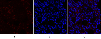
| Applications | WB;IHC;IF;ELISA |
| Other name | COL2A1; Collagen alpha-1(II) chain; Alpha-1 type II collagen |
| Size | 100μL |
| Unit price ($) | 248 |
| Human gene ID | 1280 |
| Human Swiss-Prot | P02458 |
| Source | Rabbit |
| Isotype | IgG |
| Target | Collagen II |
| Fields | >>PI3K-Akt signaling pathway;>>Focal adhesion;>>ECM-receptor interaction;>>Protein digestion and absorption;>>Human papillomavirus infection |
| Gene name | COL2A1 |
| Protein name | Collagen alpha-1(II) chain |
| Human gene link | View Human Gene |
| Human Swiss link | View Human Swiss-Prot |
| Mouse gene ID | 12824 |
| Mouse gene link | View Mouse Gene |
| Mouse Swiss-Prot | P28481 |
| Mouse Swiss link | View Mouse Swiss-Prot |
| Rat gene ID | 25412 |
| Rat gene link | View Rat Gene |
| Rat Swiss-Prot | P05539 |
| Rat Swiss link | View Rat Swiss-Prot |
| Immunogen | The antiserum was produced against synthesized peptide derived from human Collagen II. AA range:101-150 |
| Specificity | COL2A1 Polyclonal Antibody detects endogenous levels of COL2A1 protein. |
| Formulation | Liquid in PBS containing 50% glycerol, 0.5% BSA and 0.02% sodium azide. |
| Clonality | Polyclonal |
| Dilution | Western Blot: 1/500 - 1/2000. Immunohistochemistry: 1/100 - 1/300. Immunofluorescence: 1/200 - 1/1000. ELISA: 1/20000. Not yet tested in other applications. |
| Purification | The antibody was affinity-purified from rabbit antiserum by affinity-chromatography using epitope-specific immunogen. |
| Concentration | 1 mg/ml |
| Storage stability | -20°C/1 year |
| Molecular Weight (Da) | |
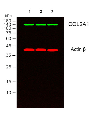
| Observed band (KD) | 140kD |
| Background | This gene encodes the alpha-1 chain of type II collagen, a fibrillar collagen found in cartilage and the vitreous humor of the eye. Mutations in this gene are associated with achondrogenesis, chondrodysplasia, early onset familial osteoarthritis, SED congenita, Langer-Saldino achondrogenesis, Kniest dysplasia, Stickler syndrome type I, and spondyloepimetaphyseal dysplasia Strudwick type. In addition, defects in processing chondrocalcin, a calcium binding protein that is the C-propeptide of this collagen molecule, are also associated with chondrodysplasia. There are two transcripts identified for this gene. [provided by RefSeq, Jul 2008], |
| Function | disease:Defects in COL2A1 are a cause of primary avascular necrosis of femoral head (ANFH) [MIM:608805]; also called ischemic necrosis of the femoral head or osteonecrosis of the femoral head. ANFH causes disability that often requires surgical intervention. Most cases are sporadic, but families in which there is an autosomal dominant inheritance of the disease have been identified. It has been estimated that 300,000 to 600,000 people in the United States have ANFH. Approximately 15,000 new cases of this common and disabling disorder are reported annually. The age at the onset is earlier than that for osteoarthritis. The diagnosis is typically made when patients are between the ages of 30 and 60 years. The clinical manifestations, such as pain on exertion, a limping gait, and a discrepancy in leg length, cause considerable disability. Moreover, nearly 10 percent of the 500,000 total-hip |
| Subcellular location | Secreted, extracellular space, extracellular matrix . |
| Expression | Isoform 2 is highly expressed in juvenile chondrocyte and low in fetal chondrocyte. |

Use collapsible tabs for more detailed information that will help customers make a purchasing decision.
Ex: Shipping and return policies, size guides, and other common questions.