 Français
 English

| ELK.No | ES2161 |
| Product name | Dematin rabbit pAb |
| Reactivity | Human;Mouse |
| Applications | WB;IHC;IF;ELISA |
| Other name | EPB49; DMT; Dematin; Erythrocyte membrane protein band 4.9 |
| Size | 50μL |
| Unit price ($) | 148 |
| Human gene ID | 2039 |
| Human Swiss-Prot | Q08495 |
| Source | Rabbit |
| Isotype | IgG |
| Target | Dematin |
| Fields | |
| Gene name | EPB49 |
| Protein name | Dematin |
| Human gene link | View Human Gene |
| Human Swiss link | View Human Swiss-Prot |
| Mouse gene ID | 13829 |
| Mouse gene link | View Mouse Gene |
| Mouse Swiss-Prot | Q9WV69 |
| Mouse Swiss link | View Mouse Swiss-Prot |
| Rat gene ID | |
| Rat gene link | |
| Rat Swiss-Prot | |
| Rat Swiss link | |
| Immunogen | The antiserum was produced against synthesized peptide derived from human Dematin. AA range:356-405 |
| Specificity | Dematin Polyclonal Antibody detects endogenous levels of Dematin protein. |
| Formulation | Liquid in PBS containing 50% glycerol, 0.5% BSA and 0.02% sodium azide. |
| Clonality | Polyclonal |
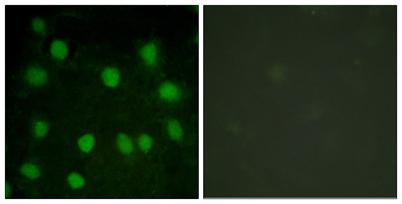
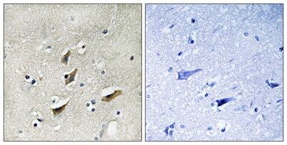
| Dilution | Western Blot: 1/500 - 1/2000. Immunohistochemistry: 1/100 - 1/300. Immunofluorescence: 1/200 - 1/1000. ELISA: 1/5000. Not yet tested in other applications. |
| Purification | The antibody was affinity-purified from rabbit antiserum by affinity-chromatography using epitope-specific immunogen. |
| Concentration | 1 mg/ml |
| Storage stability | -20°C/1 year |
| Molecular Weight (Da) | |
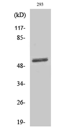
| Observed band (KD) | 50kD |
| Background | The protein encoded by this gene is an actin binding and bundling protein that plays a structural role in erythrocytes, by stabilizing and attaching the spectrin/actin cytoskeleton to the erythrocyte membrane in a phosphorylation-dependent manner. This protein contains a core domain in the N-terminus, and a headpiece domain in the C-terminus that binds F-actin. When purified from erythrocytes, this protein exists as a trimer composed of two 48 kDa polypeptides and a 52 kDa polypeptide. The different subunits arise from alternative splicing in the 3' coding region, where the headpiece domain is located. Disruption of this gene has been correlated with the autosomal dominant Marie Unna hereditary hypotrichosis disease, while loss of heterozygosity of this gene is thought to play a role in prostate cancer progression. Alternative splicing results in multiple transcript variants encoding di |
| Function | domain:Consists of a large core fragment, the amino-terminal portion, and a small headpiece, the C-terminal portion. The headpiece can bind but cannot bundle actin filaments.,domain:Contains at least two actin-binding sites, one in the headpiece domain and one in the amino-terminal portion.,function:Actin-bundling protein. May function in mitogen-activated protein kinase pathway.,PTM:Actin-bundling activity is abolished upon phosphorylation by cAMP-dependent protein kinase.,PTM:The N-terminus is blocked.,similarity:Belongs to the villin/gelsolin family.,similarity:Contains 1 HP (headpiece) domain.,subunit:Exists in solution as a trimer of two short isoforms and one long isoform linked by disulfide bonds (Probable). Interacts with RASGRF2.,tissue specificity:Heart, brain, lung, skeletal muscle, and kidney., |
| Subcellular location | Cytoplasm. Cytoplasm, cytosol. Cytoplasm, perinuclear region . Cytoplasm, cytoskeleton. Cell membrane. Membrane . Endomembrane system. Cell projection . Localized at the spectrin-actin junction of erythrocyte plasma membrane. Localized to intracellular membranes and the cytoskeletal network. Localized at intracellular membrane-bounded organelle compartment in platelets that likely represent the dense tubular network membrane. Detected at the cell membrane and at the parasitophorous vacuole in malaria-infected erythrocytes at late stages of plasmodium berghei or falciparum development. |
| Expression | Expressed in platelets (at protein level). Expressed in heart, brain, lung, skeletal muscle, and kidney. |

Use collapsible tabs for more detailed information that will help customers make a purchasing decision.
Ex: Shipping and return policies, size guides, and other common questions.