 Français
 English
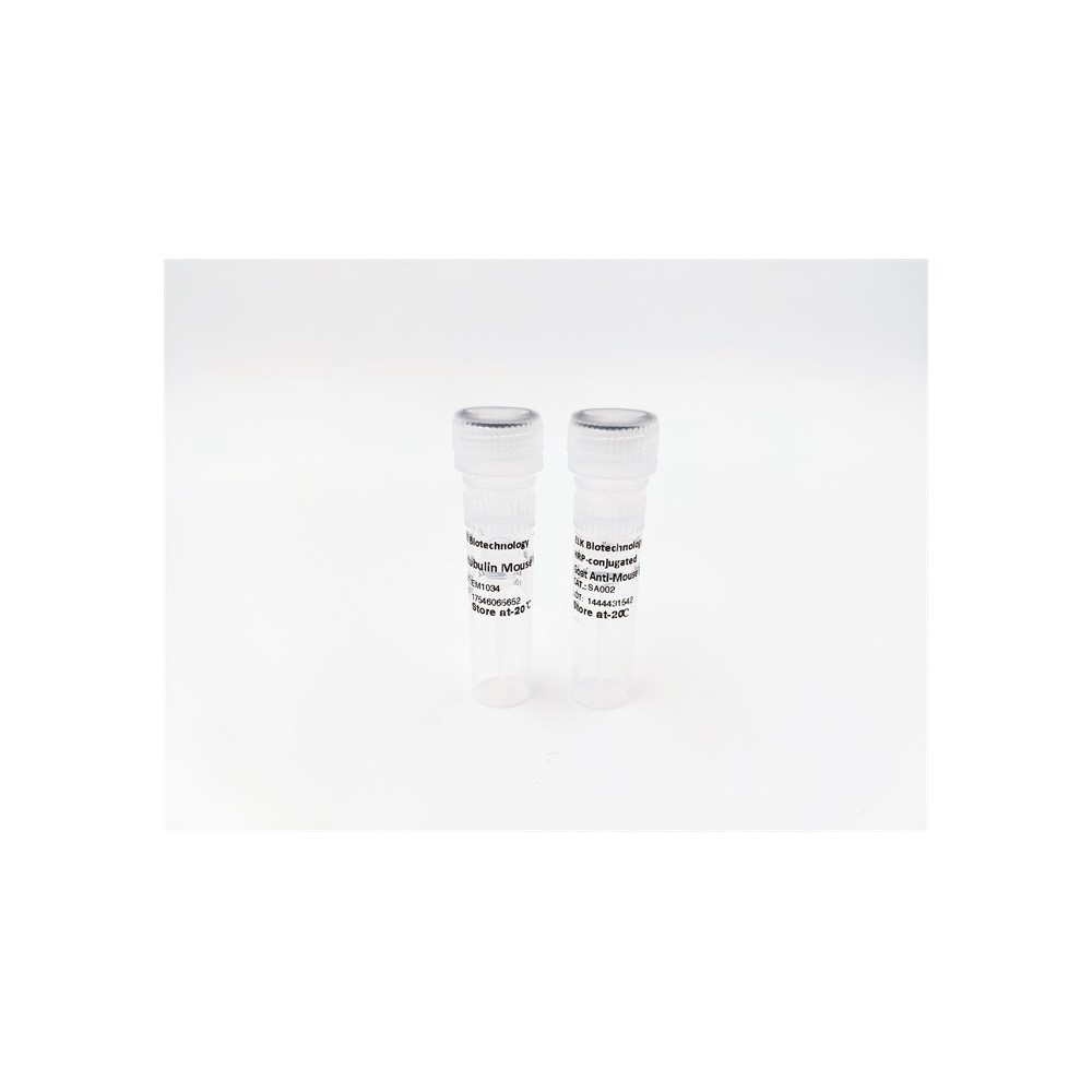

| ELK.No | ES2180 |
| Product name | DMPK rabbit pAb |
| Reactivity | Human;Rat;Mouse; |
| Applications | WB;ELISA |
| Other name | DMPK; DM1PK; MDPK; Myotonin-protein kinase; MT-PK; DM-kinase; DMK; DM1 protein kinase; DMPK; Myotonic dystrophy protein kinase |
| Size | 50μL |
| Unit price ($) | 148 |
| Human gene ID | 1760 |
| Human Swiss-Prot | Q09013 |
| Source | Rabbit |
| Isotype | IgG |
| Target | DMPK |
| Fields | |
| Gene name | DMPK |
| Protein name | Myotonin-protein kinase |
| Human gene link | View Human Gene |
| Human Swiss link | View Human Swiss-Prot |
| Mouse gene ID | |
| Mouse gene link | |
| Mouse Swiss-Prot | P54265 |
| Mouse Swiss link | View Mouse Swiss-Prot |
| Rat gene ID | |
| Rat gene link | |
| Rat Swiss-Prot | |
| Rat Swiss link | |
| Immunogen | The antiserum was produced against synthesized peptide derived from human DMPK. AA range:11-60 |
| Specificity | DMPK Polyclonal Antibody detects endogenous levels of DMPK protein. |
| Formulation | Liquid in PBS containing 50% glycerol, 0.5% BSA and 0.02% sodium azide. |
| Clonality | Polyclonal |
| Dilution | Western Blot: 1/500 - 1/2000. ELISA: 1/40000. Not yet tested in other applications. |
| Purification | The antibody was affinity-purified from rabbit antiserum by affinity-chromatography using epitope-specific immunogen. |
| Concentration | 1 mg/ml |
| Storage stability | -20°C/1 year |
| Molecular Weight (Da) | |
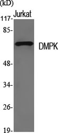
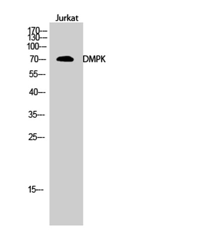
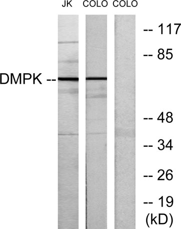
| Observed band (KD) | 70kD |
| Background | The protein encoded by this gene is a serine-threonine kinase that is closely related to other kinases that interact with members of the Rho family of small GTPases. Substrates for this enzyme include myogenin, the beta-subunit of the L-type calcium channels, and phospholemman. The 3' untranslated region of this gene contains 5-38 copies of a CTG trinucleotide repeat. Expansion of this unstable motif to 50-5,000 copies causes myotonic dystrophy type I, which increases in severity with increasing repeat element copy number. Repeat expansion is associated with condensation of local chromatin structure that disrupts the expression of genes in this region. Several alternatively spliced transcript variants of this gene have been described, but the full-length nature of some of these variants has not been determined. [provided by RefSeq, Jul 2016], |
| Function | catalytic activity:ATP + a protein = ADP + a phosphoprotein.,cofactor:Magnesium.,disease:Defects in DMPK are the cause of myotonic dystrophy 1 (DM1) [MIM:160900]; also known as Steinert disease. DM is an autosomal dominant neurodegenerative disorder characterized by myotonia, muscle wasting in the distal extremities, cataract, hypogonadism, defective endocrine functions, male baldness, and cardiac arrhythmias. DM patients show decreased levels of kinase expression inversely related to repeat length. The minimum estimated incidence is 1 in 8'000 live births. DM1 is caused by a CTG expansion in the 3'-UTR of the DMPK gene. The repeat length usually increases in successive generations, but not always.,enzyme regulation:Activated in response to G protein second messengers. Maintained in an inactive conformation by the negative autoregulatory C-terminal coiled-coil region. Coiled-coil mediate |
| Subcellular location | Endoplasmic reticulum membrane ; Single-pass type IV membrane protein ; Cytoplasmic side . Nucleus outer membrane ; Single-pass type IV membrane protein ; Cytoplasmic side . Mitochondrion outer membrane ; Single-pass type IV membrane protein . Sarcoplasmic reticulum membrane . Cell membrane . Cytoplasm, cytosol . Localizes to sarcoplasmic reticulum membranes of cardiomyocytes. .; [Isoform 1]: Mitochondrion membrane.; [Isoform 3]: Mitochondrion membrane. |
| Expression | Most isoforms are expressed in many tissues including heart, skeletal muscle, liver and brain, except for isoform 2 which is only found in the heart and skeletal muscle, and isoform 14 which is only found in the brain, with high levels in the striatum, cerebellar cortex and pons. |

Use collapsible tabs for more detailed information that will help customers make a purchasing decision.
Ex: Shipping and return policies, size guides, and other common questions.