 Français
 English

| ELK.No | ES2327 |
| Product name | FAS-L rabbit pAb |
| Reactivity | Human;Mouse;Pig |
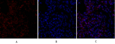
| Applications | WB;IHC;IF;ELISA |
| Other name | FASLG; APT1LG1; CD95L; FASL; TNFSF6; Tumor necrosis factor ligand superfamily member 6; Apoptosis antigen ligand; APTL; CD95 ligand; CD95-L; Fas antigen ligand; Fas ligand; FasL; CD antigen CD178 |
| Size | 50μL |
| Unit price ($) | 148 |
| Human gene ID | 356 |
| Human Swiss-Prot | P48023 |
| Source | Rabbit |
| Isotype | IgG |
| Target | FAS-L |
| Fields | >>Platinum drug resistance;>>MAPK signaling pathway;>>Ras signaling pathway;>>Cytokine-cytokine receptor interaction;>>FoxO signaling pathway;>>PI3K-Akt signaling pathway;>>Apoptosis;>>Necroptosis;>>Natural killer cell mediated cytotoxicity;>>Neurotrophin signaling pathway;>>Non-alcoholic fatty liver disease;>>Alcoholic liver disease;>>Type I diabetes mellitus;>>Pathways of neurodegeneration - multiple diseases;>>Pathogenic Escherichia coli infection;>>Chagas disease;>>African trypanosomiasis;>>Hepatitis C;>>Hepatitis B;>>Measles;>>Human cytomegalovirus infection;>>Influenza A;>>Human papillomavirus infection;>>Herpes simplex virus 1 infection;>>Human immunodeficiency virus 1 infection;>>Pathways in cancer;>>Proteoglycans in cancer;>>Autoimmune thyroid disease;>>Allograft rejection;>>Graft-versus-host disease;>>Lipid and atherosclerosis |
| Gene name | FASLG |
| Protein name | Tumor necrosis factor ligand superfamily member 6 |
| Human gene link | View Human Gene |
| Human Swiss link | View Human Swiss-Prot |
| Mouse gene ID | 14103 |
| Mouse gene link | View Mouse Gene |
| Mouse Swiss-Prot | P41047 |
| Mouse Swiss link | View Mouse Swiss-Prot |
| Rat gene ID | |
| Rat gene link | |
| Rat Swiss-Prot | |
| Rat Swiss link | |
| Immunogen | The antiserum was produced against synthesized peptide derived from human FAS ligand. AA range:101-150 |
| Specificity | FAS-L Polyclonal Antibody detects endogenous levels of FAS-L protein. |
| Formulation | Liquid in PBS containing 50% glycerol, 0.5% BSA and 0.02% sodium azide. |
| Clonality | Polyclonal |
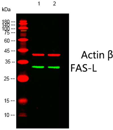
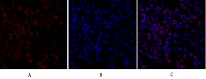
| Dilution | Western Blot: 1/500 - 1/2000. Immunohistochemistry: 1/100 - 1/300. Immunofluorescence: 1/200 - 1/1000. ELISA: 1/40000. Not yet tested in other applications. |
| Purification | The antibody was affinity-purified from rabbit antiserum by affinity-chromatography using epitope-specific immunogen. |
| Concentration | 1 mg/ml |
| Storage stability | -20°C/1 year |
| Molecular Weight (Da) | |
| Observed band (KD) | 33kD |
| Background | This gene is a member of the tumor necrosis factor superfamily. The primary function of the encoded transmembrane protein is the induction of apoptosis triggered by binding to FAS. The FAS/FASLG signaling pathway is essential for immune system regulation, including activation-induced cell death (AICD) of T cells and cytotoxic T lymphocyte induced cell death. It has also been implicated in the progression of several cancers. Defects in this gene may be related to some cases of systemic lupus erythematosus (SLE). Alternatively spliced transcript variants have been described. [provided by RefSeq, Nov 2014], |
| Function | disease:Defects in FASLG are the cause of autoimmune lymphoproliferative syndrome type 1B (ALPS1B) [MIM:601859]; also known as Canale-Smith syndrome (CSS). ALPS is a childhood syndrome involving hemolytic anemia and thrombocytopenia with massive lymphadenopathy and splenomegaly.,function:Cytokine that binds to TNFRSF6/FAS, a receptor that transduces the apoptotic signal into cells. May be involved in cytotoxic T-cell mediated apoptosis and in T-cell development. TNFRSF6/FAS-mediated apoptosis may have a role in the induction of peripheral tolerance, in the antigen-stimulated suicide of mature T-cells, or both. Binding to the decoy receptor TNFRSF6B/DcR3 modulates its effects.,online information:FAS-ligand entry,online information:FASLG mutation db,PTM:N-glycosylated.,PTM:The soluble form derives from the membrane form by proteolytic processing.,similarity:Belongs to the tumor necrosis fa |
| Subcellular location | Cell membrane ; Single-pass type II membrane protein . Cytoplasmic vesicle lumen . Lysosome lumen . Is internalized into multivesicular bodies of secretory lysosomes after phosphorylation by FGR and monoubiquitination (PubMed:17164290). Colocalizes with the SPPL2A protease at the cell membrane (PubMed:17557115). .; [Tumor necrosis factor ligand superfamily member 6, soluble form]: Secreted . May be released into the extracellular fluid by cleavage from the cell surface. .; [FasL intracellular domain]: Nucleus . The FasL ICD cytoplasmic form is translocated into the nucleus. . |
| Expression | Blood,Leukocyte,Spleen, |

Use collapsible tabs for more detailed information that will help customers make a purchasing decision.
Ex: Shipping and return policies, size guides, and other common questions.