 Français
 English
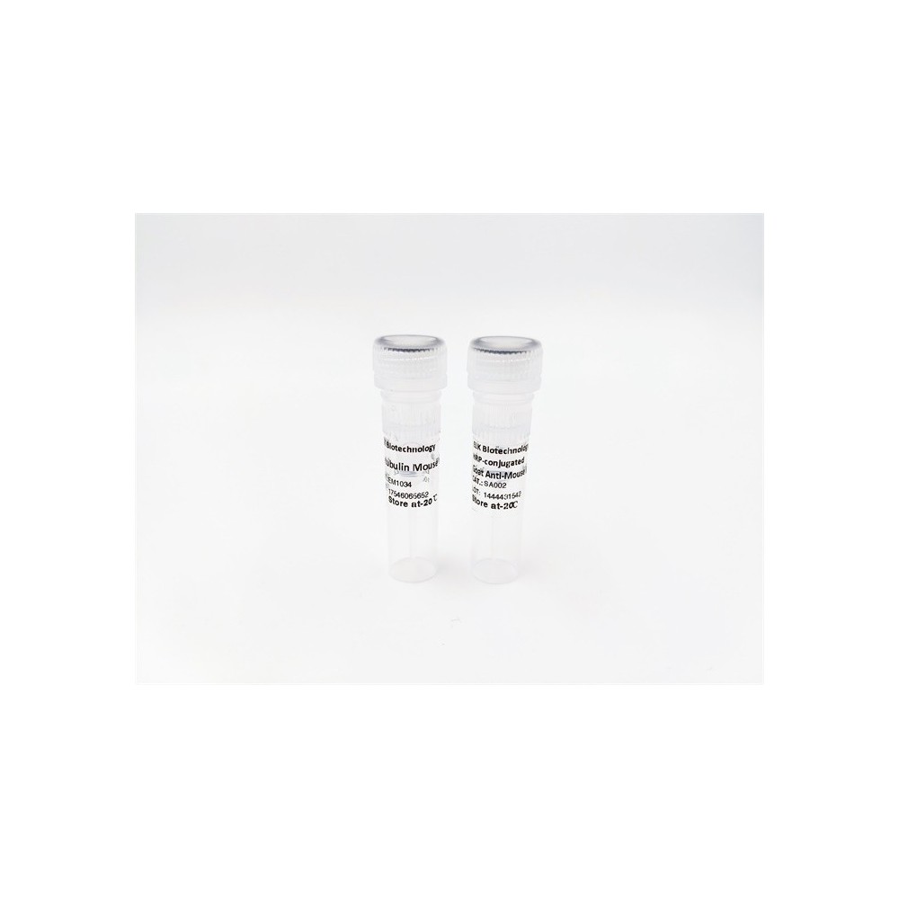

| ELK.No | ES2356 |
| Product name | Fnk rabbit pAb |
| Reactivity | Human;Mouse;Rat |
| Applications | WB;ELISA |
| Other name | PLK3; CNK; FNK; PRK; Serine/threonine-protein kinase PLK3; Cytokine-inducible serine/threonine-protein kinase; FGF-inducible kinase; Polo-like kinase 3; PLK-3; Proliferation-related kinase |
| Size | 50μL |
| Unit price ($) | 148 |
| Human gene ID | 1263 |
| Human Swiss-Prot | Q9H4B4 |
| Source | Rabbit |
| Isotype | IgG |
| Target | Fnk |
| Fields | >>FoxO signaling pathway;>>C-type lectin receptor signaling pathway;>>Tuberculosis |
| Gene name | PLK3 |
| Protein name | Serine/threonine-protein kinase PLK3 |
| Human gene link | View Human Gene |
| Human Swiss link | View Human Swiss-Prot |
| Mouse gene ID | |
| Mouse gene link | |
| Mouse Swiss-Prot | Q60806 |
| Mouse Swiss link | View Mouse Swiss-Prot |
| Rat gene ID | 58936 |
| Rat gene link | View Rat Gene |
| Rat Swiss-Prot | Q9R011 |
| Rat Swiss link | View Rat Swiss-Prot |
| Immunogen | The antiserum was produced against synthesized peptide derived from human PLK3. AA range:231-280 |
| Specificity | Fnk Polyclonal Antibody detects endogenous levels of Fnk protein. |
| Formulation | Liquid in PBS containing 50% glycerol, 0.5% BSA and 0.02% sodium azide. |
| Clonality | Polyclonal |
| Dilution | Western Blot: 1/500 - 1/2000. ELISA: 1/10000. Not yet tested in other applications. |
| Purification | The antibody was affinity-purified from rabbit antiserum by affinity-chromatography using epitope-specific immunogen. |
| Concentration | 1 mg/ml |
| Storage stability | -20°C/1 year |
| Molecular Weight (Da) | |
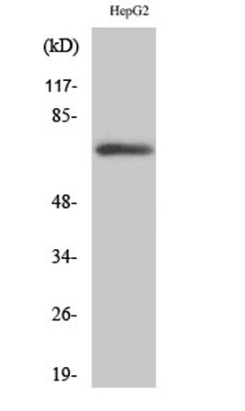
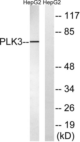
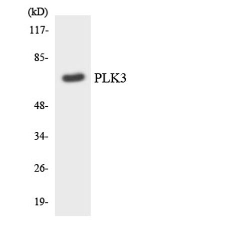
| Observed band (KD) | 70kD |
| Background | The protein encoded by this gene is a member of the highly conserved polo-like kinase family of serine/threonine kinases. Members of this family are characterized by an amino-terminal kinase domain and a carboxy-terminal bipartite polo box domain that functions as a substrate-binding motif and a cellular localization signal. Polo-like kinases are important regulators of cell cycle progression. This gene has also been implicated in stress responses and double-strand break repair. In human cell lines, this protein is reported to associate with centrosomes in a microtubule-dependent manner, and during mitosis, the protein becomes localized to the mitotic apparatus. Expression of a kinase-defective mutant results in abnormal cell morphology caused by changes in microtubule dynamics and mitotic arrest followed by apoptosis. [provided by RefSeq, Sep 2015], |
| Function | catalytic activity:ATP + a protein = ADP + a phosphoprotein.,function:Serine/threonine protein kinase involved in regulating M phase functions during the cell cycle. May also be part of the signaling network controlling cellular adhesion. In vitro, is able to phosphorylate CDC25C and casein.,induction:Cytokine and cellular adhesion trigger FNK induction.,PTM:Phosphorylated as cells enter mitosis and dephosphorylated as cells exit mitosis.,similarity:Belongs to the protein kinase superfamily.,similarity:Belongs to the protein kinase superfamily. Ser/Thr protein kinase family. CDC5/Polo subfamily.,similarity:Contains 1 protein kinase domain.,similarity:Contains 2 POLO box domains.,subunit:Binds to the calcium/integrin-binding protein (CIB). This interaction probably occurs via the POLO-box domain.,tissue specificity:Transcripts are highly detected in placenta, lung, followed by skeletal mu |
| Subcellular location | Cytoplasm. Nucleus. Nucleus, nucleolus. Golgi apparatus. Cytoplasm, cytoskeleton, microtubule organizing center, centrosome. Translocates to the nucleus upon cisplatin treatment. Localizes to the Golgi apparatus during interphase. According to a report, PLK3 localizes only in the nucleolus and not in the centrosome, or in any other location in the cytoplasm (PubMed:17264206). The discrepancies in results may be explained by the PLK3 antibody specificity, by cell line-specific expression or post-translational modifications. . |
| Expression | Transcripts are highly detected in placenta, lung, followed by skeletal muscle, heart, pancreas, ovaries and kidney and weakly detected in liver and brain. May have a short half-live. In cells of hematopoietic origin, strongly and exclusively detected in terminally differentiated macrophages. Transcript expression appears to be down-regulated in primary lung tumor. |

Use collapsible tabs for more detailed information that will help customers make a purchasing decision.
Ex: Shipping and return policies, size guides, and other common questions.