 Français
 English

| ELK.No | ES2459 |
| Product name | GPR176 rabbit pAb |
| Reactivity | Human;Mouse;Rat |
| Applications | WB;IF;ELISA |
| Other name | GPR176; Probable G-protein coupled receptor 176; HB-954 |
| Size | 50μL |
| Unit price ($) | 148 |
| Human gene ID | 11245 |
| Human Swiss-Prot | Q14439 |
| Source | Rabbit |
| Isotype | IgG |
| Target | GPR176 |
| Fields | |
| Gene name | GPR176 |
| Protein name | Probable G-protein coupled receptor 176 |
| Human gene link | View Human Gene |
| Human Swiss link | View Human Swiss-Prot |
| Mouse gene ID | 381413 |
| Mouse gene link | View Mouse Gene |
| Mouse Swiss-Prot | Q80WT4 |
| Mouse Swiss link | View Mouse Swiss-Prot |
| Rat gene ID | 117257 |
| Rat gene link | View Rat Gene |
| Rat Swiss-Prot | Q64017 |
| Rat Swiss link | View Rat Swiss-Prot |
| Immunogen | The antiserum was produced against synthesized peptide derived from human GPR176. AA range:466-515 |
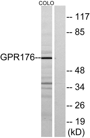
| Specificity | GPR176 Polyclonal Antibody detects endogenous levels of GPR176 protein. |
| Formulation | Liquid in PBS containing 50% glycerol, 0.5% BSA and 0.02% sodium azide. |
| Clonality | Polyclonal |
| Dilution | Western Blot: 1/500 - 1/2000. Immunofluorescence: 1/200 - 1/1000. ELISA: 1/10000. Not yet tested in other applications. |
| Purification | The antibody was affinity-purified from rabbit antiserum by affinity-chromatography using epitope-specific immunogen. |
| Concentration | 1 mg/ml |
| Storage stability | -20°C/1 year |
| Molecular Weight (Da) | |
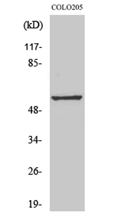
| Observed band (KD) | 57kD |
| Background | Members of the G protein-coupled receptor family, such as GPR176, are cell surface receptors involved in responses to hormones, growth factors, and neurotransmitters (Hata et al., 1995 [PubMed 7893747]).[supplied by OMIM, Jul 2008], |
| Function | function:Orphan receptor.,similarity:Belongs to the G-protein coupled receptor 1 family., |
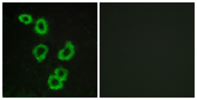
| Subcellular location | Cell membrane ; Multi-pass membrane protein . |
| Expression | Fetal brain,Ovary, |

Use collapsible tabs for more detailed information that will help customers make a purchasing decision.
Ex: Shipping and return policies, size guides, and other common questions.