 Français
 English

| ELK.No | ES2660 |
| Product name | JIP-3 rabbit pAb |
| Reactivity | Human;Mouse |
| Applications | WB;IHC;IF;ELISA |
| Other name | MAPK8IP3; JIP3; KIAA1066; C-Jun-amino-terminal kinase-interacting protein 3; JIP-3; JNK-interacting protein 3; JNK MAP kinase scaffold protein 3; Mitogen-activated protein kinase 8-interacting protein 3 |
| Size | 100μL |
| Unit price ($) | 248 |
| Human gene ID | 23162 |
| Human Swiss-Prot | Q9UPT6 |
| Source | Rabbit |
| Isotype | IgG |
| Target | JIP-3 |
| Fields | >>MAPK signaling pathway |
| Gene name | MAPK8IP3 |
| Protein name | C-Jun-amino-terminal kinase-interacting protein 3 |
| Human gene link | View Human Gene |
| Human Swiss link | View Human Swiss-Prot |
| Mouse gene ID | 30957 |
| Mouse gene link | View Mouse Gene |
| Mouse Swiss-Prot | Q9ESN9 |
| Mouse Swiss link | View Mouse Swiss-Prot |
| Rat gene ID | |
| Rat gene link | |
| Rat Swiss-Prot | |
| Rat Swiss link | |
| Immunogen | The antiserum was produced against synthesized peptide derived from human JIP3. AA range:621-670 |
| Specificity | JIP-3 Polyclonal Antibody detects endogenous levels of JIP-3 protein. |
| Formulation | Liquid in PBS containing 50% glycerol, 0.5% BSA and 0.02% sodium azide. |
| Clonality | Polyclonal |
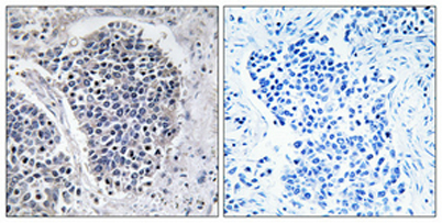
| Dilution | Western Blot: 1/500 - 1/2000. Immunohistochemistry: 1/100 - 1/300. Immunofluorescence: 1/200 - 1/1000. ELISA: 1/20000. Not yet tested in other applications. |
| Purification | The antibody was affinity-purified from rabbit antiserum by affinity-chromatography using epitope-specific immunogen. |
| Concentration | 1 mg/ml |
| Storage stability | -20°C/1 year |
| Molecular Weight (Da) | |
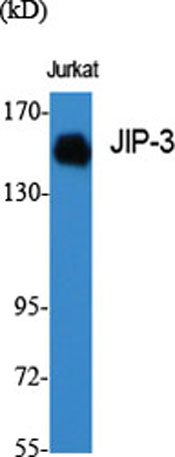
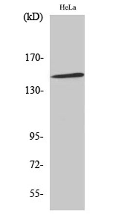
| Observed band (KD) | 147kD |
| Background | The protein encoded by this gene shares similarity with the product of Drosophila syd gene, required for the functional interaction of kinesin I with axonal cargo. Studies of the similar gene in mouse suggested that this protein may interact with, and regulate the activity of numerous protein kinases of the JNK signaling pathway, and thus function as a scaffold protein in neuronal cells. The C. elegans counterpart of this gene is found to regulate synaptic vesicle transport possibly by integrating JNK signaling and kinesin-1 transport. Several alternatively spliced transcript variants of this gene have been described, but the full-length nature of some of these variants has not been determined. [provided by RefSeq, Jul 2008], |
| Function | function:The JNK-interacting protein (JIP) group of scaffold proteins selectively mediates JNK signaling by aggregating specific components of the MAPK cascade to form a functional JNK signaling module. May function as a regulator of vesicle transport, through interations with the JNK-signaling components and motor proteins.,PTM:Phosphorylated upon DNA damage, probably by ATM or ATR.,similarity:Belongs to the JIP scaffold family.,subunit:Forms homo- or heterooligomeric complexes. The central region of MAPK8IP3 interacts with the C-terminal of MAPK8IP2 but not MAPK8IP1. Binds specific components of the JNK signaling pathway namely MAPK8, MAPK9 and MAPK10 to the N-terminal region, MAP2K4 and MAP2K7 to the central region and MAP3K11 to the C-terminal region. Binds the TPR motif-containing C-terminal of kinesin light chain, KLC1. Pre-assembled MAPK8IP1 scaffolding complexes are then transpor |
| Subcellular location | Cytoplasm . Golgi apparatus . Cytoplasmic vesicle . Cell projection, growth cone . Cell projection, axon . Cell projection, dendrite . Cytoplasm, perinuclear region . Localized in the soma and growth cones of differentiated neurites and the Golgi and vesicles of the early secretory compartment of epithelial cells. KIF5A/B/C-mediated transportation to axon tips is essential for its function in enhancing neuronal axon elongation. . |
| Expression | Brain,Epithelium,Melanoma,Spleen, |

Use collapsible tabs for more detailed information that will help customers make a purchasing decision.
Ex: Shipping and return policies, size guides, and other common questions.