 Français
 English
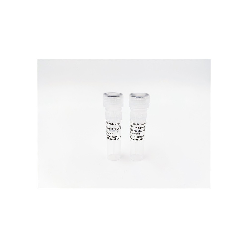

| ELK.No | ES2704 |
| Product name | LATS1/2 rabbit pAb |
| Reactivity | Human;Mouse |
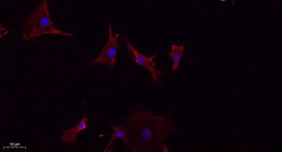
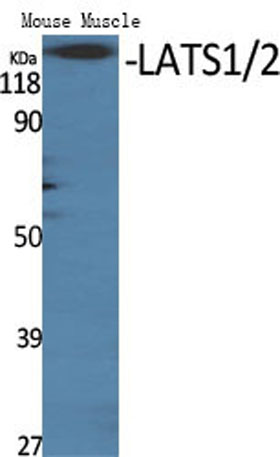
| Applications | WB;IHC;IF;ELISA |
| Other name | LATS1; WARTS; Serine/threonine-protein kinase LATS1; Large tumor suppressor homolog 1; WARTS protein kinase; h-warts; LATS2; KPM; Serine/threonine-protein kinase LATS2; Kinase phosphorylated during mitosis protein; Large tumor suppressor ho |
| Size | 50μL |
| Unit price ($) | 148 |
| Human gene ID | 9113/26524 |
| Human Swiss-Prot | O95835/Q9NRM7 |
| Source | Rabbit |
| Isotype | IgG |
| Target | LATS1/2 |
| Fields | >>Hippo signaling pathway;>>Hippo signaling pathway - multiple species |
| Gene name | LATS1/LATS2 |
| Protein name | Serine/threonine-protein kinase LATS1/2 |
| Human gene link | View Human Gene |
| Human Swiss link | View Human Swiss-Prot |
| Mouse gene ID | 16798/50523 |
| Mouse gene link | View Mouse Gene |
| Mouse Swiss-Prot | |
| Mouse Swiss link | |
| Rat gene ID | |
| Rat gene link | |
| Rat Swiss-Prot | |
| Rat Swiss link | |
| Immunogen | The antiserum was produced against synthesized peptide derived from human LATS1/2. AA range:1041-1090 |
| Specificity | LATS1/2 Polyclonal Antibody detects endogenous levels of LATS1/2 protein. |
| Formulation | Liquid in PBS containing 50% glycerol, 0.5% BSA and 0.02% sodium azide. |
| Clonality | Polyclonal |
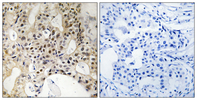
| Dilution | Western Blot: 1/500 - 1/2000. IHC-p: 1:100-300 ELISA: 1/20000. IF 1:100-300 Not yet tested in other applications. |
| Purification | The antibody was affinity-purified from rabbit antiserum by affinity-chromatography using epitope-specific immunogen. |
| Concentration | 1 mg/ml |
| Storage stability | -20°C/1 year |
| Molecular Weight (Da) | |
| Observed band (KD) | 130-140kD |
| Background | The protein encoded by this gene is a putative serine/threonine kinase that localizes to the mitotic apparatus and complexes with cell cycle controller CDC2 kinase in early mitosis. The protein is phosphorylated in a cell-cycle dependent manner, with late prophase phosphorylation remaining through metaphase. The N-terminal region of the protein binds CDC2 to form a complex showing reduced H1 histone kinase activity, indicating a role as a negative regulator of CDC2/cyclin A. In addition, the C-terminal kinase domain binds to its own N-terminal region, suggesting potential negative regulation through interference with complex formation via intramolecular binding. Biochemical and genetic data suggest a role as a tumor suppressor. This is supported by studies in knockout mice showing development of soft-tissue sarcomas, ovarian stromal cell tumors and a high sensitivity to carcinogenic treatmen |
| Function | catalytic activity:ATP + a protein = ADP + a phosphoprotein.,cofactor:Magnesium.,function:Tumor suppressor which plays a critical role in maintenance of ploidy through its actions in both mitotic progression and the G1 tetraploidy checkpoint. Negatively regulates G2/M transition by down-regulating CDC2 kinase activity. Involved in the control of p53 expression. Affects cytokinesis by regulating actin polymerization through negative modulation of LIMK1. May also play a role in endocrine function.,PTM:Autophosphorylated and phosphorylated during M-phase of the cell cycle. Phosphorylated by STK3 at Ser-909 and Thr-1079, which results in its activation. Phosphorylated upon DNA damage, probably by ATM or ATR.,similarity:Belongs to the protein kinase superfamily. AGC Ser/Thr protein kinase family.,similarity:Contains 1 AGC-kinase C-terminal domain.,similarity:Contains 1 protein kinase domain., |
| Subcellular location | Cytoplasm, cytoskeleton, microtubule organizing center, centrosome . Cytoplasm, cytoskeleton, spindle . Midbody . Cytoplasm, cytoskeleton, microtubule organizing center, spindle pole body . Localizes to the centrosomes throughout interphase but migrates to the mitotic apparatus, including spindle pole bodies, mitotic spindle, and midbody, during mitosis. . |
| Expression | Expressed in all adult tissues examined except for lung and kidney. |

Use collapsible tabs for more detailed information that will help customers make a purchasing decision.
Ex: Shipping and return policies, size guides, and other common questions.