 Français
 English

| ELK.No | ES2798 |
| Product name | ML-IAP rabbit pAb |
| Reactivity | Human;Rat;Mouse; |
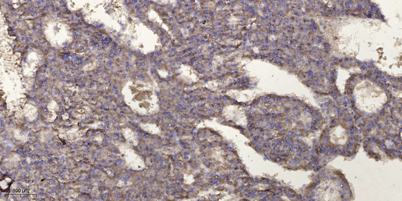
| Applications | WB;IHC |
| Other name | BIRC7; KIAP; LIVIN; MLIAP; RNF50; Baculoviral IAP repeat-containing protein 7; Kidney inhibitor of apoptosis protein; KIAP; Livin; Melanoma inhibitor of apoptosis protein; ML-IAP; RING finger protein 50 |
| Size | 50μL |
| Unit price ($) | 148 |
| Human gene ID | 79444 |
| Human Swiss-Prot | Q96CA5 |
| Source | Rabbit |
| Isotype | IgG |
| Target | ML-IAP |
| Fields | >>Ubiquitin mediated proteolysis;>>Apoptosis - multiple species;>>Toxoplasmosis;>>Pathways in cancer;>>Small cell lung cancer |
| Gene name | BIRC7 |
| Protein name | Baculoviral IAP repeat-containing protein 7 |
| Human gene link | View Human Gene |
| Human Swiss link | View Human Swiss-Prot |
| Mouse gene ID | |
| Mouse gene link | |
| Mouse Swiss-Prot | A2AWP0 |
| Mouse Swiss link | View Mouse Swiss-Prot |
| Rat gene ID | |
| Rat gene link | |
| Rat Swiss-Prot | |
| Rat Swiss link | |
| Immunogen | The antiserum was produced against synthesized peptide derived from human ML-IAP. AA range:162-211 |
| Specificity | ML-IAP Polyclonal Antibody detects endogenous levels of ML-IAP protein. |
| Formulation | Liquid in PBS containing 50% glycerol, 0.5% BSA and 0.02% sodium azide. |
| Clonality | Polyclonal |
| Dilution | WB 1:500-2000;IHC-p 1:50-300 |
| Purification | The antibody was affinity-purified from rabbit antiserum by affinity-chromatography using epitope-specific immunogen. |
| Concentration | 1 mg/ml |
| Storage stability | -20°C/1 year |
| Molecular Weight (Da) | |
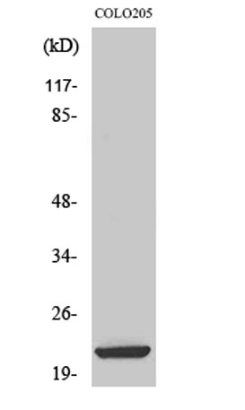
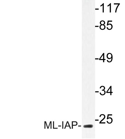
| Observed band (KD) | 21kD |
| Background | This gene encodes a member of the inhibitor of apoptosis protein (IAP) family, and contains a single copy of a baculovirus IAP repeat (BIR) as well as a RING-type zinc finger domain. The BIR domain is essential for inhibitory activity and interacts with caspases, while the RING finger domain sometimes enhances antiapoptotic activity but does not inhibit apoptosis alone. Elevated levels of the encoded protein may be associated with cancer progression and play a role in chemotherapy sensitivity. Alternative splicing results in multiple transcript variants [provided by RefSeq, Jul 2013], |
| Function | function:Protects against apoptosis induced by TNF or by chemical agents such as adriamycin, etoposide or staurosporine. Suppression of apoptosis is mediated by activation of MAPK8/JNK1, and possibly also of MAPK9/JNK2. This activation depends on TAB1 and NR2C2/TAK1. In vitro, inhibits caspase-3 and proteolytic activation of pro-caspase-9. Isoform 1 blocks staurosporine-induced apoptosis and isoform 2 blocks etoposide-induced apoptosis.,similarity:Belongs to the IAP family.,similarity:Contains 1 BIR repeat.,similarity:Contains 1 RING-type zinc finger.,subcellular location:Nuclear, and in a filamentous pattern throughout the cytoplasm.,subunit:Binds to caspase-9. Interaction with SMAC via the BIR domain disrupts binding to caspase-9 and apoptotic suppressor activity. Interacts with TAB1. In vitro, interacts with caspase-3 and caspase-7 via its BIR domain.,tissue specificity:Very low level |
| Subcellular location | Nucleus . Cytoplasm . Golgi apparatus . Nuclear, and in a filamentous pattern throughout the cytoplasm. Full-length livin is detected exclusively in the cytoplasm, whereas the truncated form (tLivin) is found in the peri-nuclear region with marked localization to the Golgi apparatus; the accumulation of tLivin in the nucleus shows positive correlation with the increase in apoptosis. |
| Expression | Isoform 1 and isoform 2 are expressed at very low levels or not detectable in most adult tissues. Detected in adult heart, placenta, lung, lymph node, spleen and ovary, and in several carcinoma cell lines. Isoform 2 is detected in fetal kidney, heart and spleen, and at lower levels in adult brain, skeletal muscle and peripheral blood leukocytes. |

Use collapsible tabs for more detailed information that will help customers make a purchasing decision.
Ex: Shipping and return policies, size guides, and other common questions.