 Français
 English

| ELK.No | ES2816 |
| Product name | MPP9 rabbit pAb |
| Reactivity | Human;Rat;Mouse; |
| Applications | WB;IHC |
| Other name | MPHOSPH9; MPP9; M-phase phosphoprotein 9 |
| Size | 50μL |
| Unit price ($) | 148 |
| Human gene ID | 10198 |
| Human Swiss-Prot | Q99550 |
| Source | Rabbit |
| Isotype | IgG |
| Target | MPP9 |
| Fields | |
| Gene name | MPHOSPH9 |
| Protein name | M-phase phosphoprotein 9 |
| Human gene link | View Human Gene |
| Human Swiss link | View Human Swiss-Prot |
| Mouse gene ID | |
| Mouse gene link | |
| Mouse Swiss-Prot | A6H5Y1 |
| Mouse Swiss link | View Mouse Swiss-Prot |
| Rat gene ID | |
| Rat gene link | |
| Rat Swiss-Prot | |
| Rat Swiss link | |
| Immunogen | The antiserum was produced against synthesized peptide derived from human MPHOSPH9. AA range:539-588 |
| Specificity | MPP9 Polyclonal Antibody detects endogenous levels of MPP9 protein. |
| Formulation | Liquid in PBS containing 50% glycerol, 0.5% BSA and 0.02% sodium azide. |
| Clonality | Polyclonal |
| Dilution | WB 1:500-2000;IHC-p 1:50-300 |
| Purification | The antibody was affinity-purified from rabbit antiserum by affinity-chromatography using epitope-specific immunogen. |
| Concentration | 1 mg/ml |
| Storage stability | -20°C/1 year |
| Molecular Weight (Da) | |
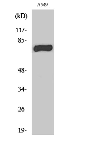
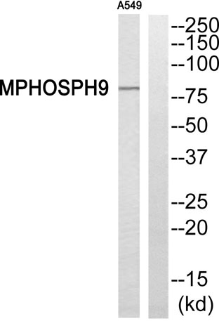
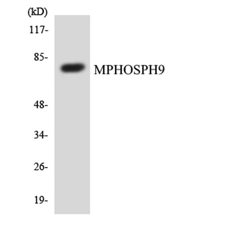
| Observed band (KD) | 78kD |
| Background | PTM:Phosphorylated in M (mitotic) phase., |
| Function | PTM:Phosphorylated in M (mitotic) phase., |
| Subcellular location | Cytoplasm, cytoskeleton, microtubule organizing center, centrosome, centriole . Golgi apparatus membrane ; Peripheral membrane protein . Cytoplasm, cytoskeleton, microtubule organizing center, centrosome . Localizes to the distal and proximal end of centriole pairs in duplicated centrosomes. In ciliated cells, localizes to the distal and proximal end of daughter centriole and proximal of the mother centriole but not in the distal end of the mother centriole (PubMed:21399614). Recruited by KIF24 to the distal end of mother centriole where it forms a ring-like structure (PubMed:30375385). . |
| Expression | Lymphoblast,Tongue, |

Use collapsible tabs for more detailed information that will help customers make a purchasing decision.
Ex: Shipping and return policies, size guides, and other common questions.