 Français
 English

| ELK.No | ES2879 |
| Product name | Myomesin-2 rabbit pAb |
| Reactivity | Human;Mouse;Monkey |
| Applications | WB;ELISA;IHC |
| Other name | MYOM2; Myomesin-2; 165 kDa connectin-associated protein; 165 kDa titin-associated protein; M-protein; Myomesin family member 2 |
| Size | 100μL |
| Unit price ($) | 248 |
| Human gene ID | 9172 |
| Human Swiss-Prot | P54296 |
| Source | Rabbit |
| Isotype | IgG |
| Target | Myomesin-2 |
| Fields | |
| Gene name | MYOM2 |
| Protein name | Myomesin-2 |
| Human gene link | View Human Gene |
| Human Swiss link | View Human Swiss-Prot |
| Mouse gene ID | |
| Mouse gene link | |
| Mouse Swiss-Prot | |
| Mouse Swiss link | |
| Rat gene ID | |
| Rat gene link | |
| Rat Swiss-Prot | |
| Rat Swiss link | |
| Immunogen | The antiserum was produced against synthesized peptide derived from human MYOM2. AA range:612-661 |
| Specificity | Myomesin-2 Polyclonal Antibody detects endogenous levels of Myomesin-2 protein. |
| Formulation | Liquid in PBS containing 50% glycerol, 0.5% BSA and 0.02% sodium azide. |
| Clonality | Polyclonal |
| Dilution | WB 1:500-2000;IHC-p 1:50-300; ELISA 2000-20000 |
| Purification | The antibody was affinity-purified from rabbit antiserum by affinity-chromatography using epitope-specific immunogen. |
| Concentration | 1 mg/ml |
| Storage stability | -20°C/1 year |
| Molecular Weight (Da) | |
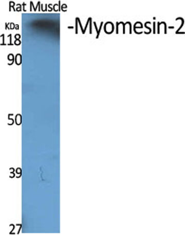
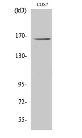
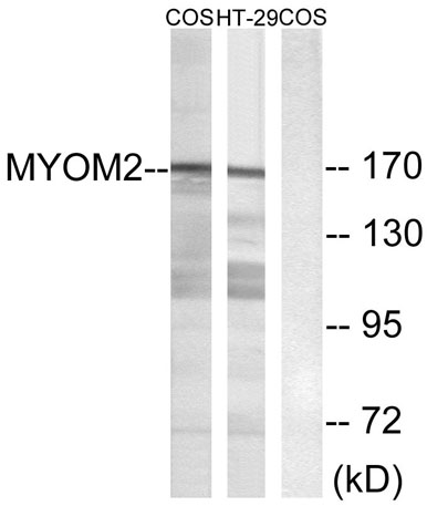
| Observed band (KD) | 165kD |
| Background | The giant protein titin, together with its associated proteins, interconnects the major structure of sarcomeres, the M bands and Z discs. The C-terminal end of the titin string extends into the M line, where it binds tightly to M-band constituents of apparent molecular masses of 190 kD and 165 kD. The predicted MYOM2 protein contains 1,465 amino acids. Like MYOM1, MYOM2 has a unique N-terminal domain followed by 12 repeat domains with strong homology to either fibronectin type III or immunoglobulin C2 domains. Protein sequence comparisons suggested that the MYOM2 protein and bovine M protein are identical. [provided by RefSeq, Jul 2008], |
| Function | function:Major component of the vertebrate myofibrillar M band. Binds myosin, titin, and light meromyosin. This binding is dose dependent.,similarity:Contains 5 fibronectin type-III domains.,similarity:Contains 5 Ig-like C2-type (immunoglobulin-like) domains.,subunit:Interacts with TTN/titin., |
| Subcellular location | Cytoplasm, myofibril, sarcomere, M line . |
| Expression | PNS,Skeletal muscle, |

Use collapsible tabs for more detailed information that will help customers make a purchasing decision.
Ex: Shipping and return policies, size guides, and other common questions.