 Français
 English
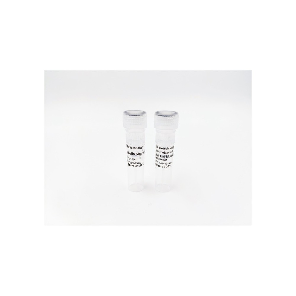

| ELK.No | ES3205 |
| Product name | PIASx rabbit pAb |
| Reactivity | Human;Mouse;Rat;Monkey |
| Applications | WB;ELISA;IHC |
| Other name | PIAS2; PIASX; E3 SUMO-protein ligase PIAS2; Androgen receptor-interacting protein 3; ARIP3; DAB2-interacting protein; DIP; Msx-interacting zinc finger protein; Miz1; PIAS-NY protein; Protein inhibitor of activated STAT x; Protein inhibitor |
| Size | 50μL |
| Unit price ($) | 148 |
| Human gene ID | 9063 |
| Human Swiss-Prot | O75928 |
| Source | Rabbit |
| Isotype | IgG |
| Target | PIAS2 |
| Fields | >>Ubiquitin mediated proteolysis;>>JAK-STAT signaling pathway |
| Gene name | PIAS2 |
| Protein name | E3 SUMO-protein ligase PIAS2 |
| Human gene link | View Human Gene |
| Human Swiss link | View Human Swiss-Prot |
| Mouse gene ID | 17344 |
| Mouse gene link | View Mouse Gene |
| Mouse Swiss-Prot | Q8C5D8 |
| Mouse Swiss link | View Mouse Swiss-Prot |
| Rat gene ID | 83422 |
| Rat gene link | View Rat Gene |
| Rat Swiss-Prot | Q6AZ28 |
| Rat Swiss link | View Rat Swiss-Prot |
| Immunogen | The antiserum was produced against synthesized peptide derived from human PIAS2. AA range:10-59 |
| Specificity | PIASx Polyclonal Antibody detects endogenous levels of PIASx protein. |
| Formulation | Liquid in PBS containing 50% glycerol, 0.5% BSA and 0.02% sodium azide. |
| Clonality | Polyclonal |
| Dilution | WB 1:500-2000;IHC-p 1:50-300; ELISA 2000-20000 |
| Purification | The antibody was affinity-purified from rabbit antiserum by affinity-chromatography using epitope-specific immunogen. |
| Concentration | 1 mg/ml |
| Storage stability | -20°C/1 year |
| Molecular Weight (Da) | |
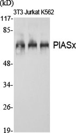
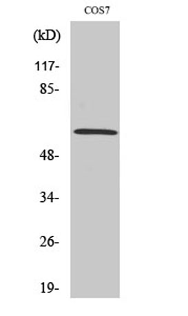
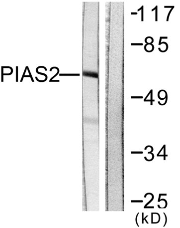
| Observed band (KD) | 68kD |
| Background | This gene encodes a member of the protein inhibitor of activated STAT (PIAS) family. PIAS proteins function as SUMO E3 ligases and play important roles in many cellular processes by mediating the sumoylation of target proteins. Alternatively spliced transcript variants encoding multiple isoforms have been observed for this gene. Isoforms of the encoded protein enhance the sumoylation of specific target proteins including the p53 tumor suppressor protein, c-Jun, and the androgen receptor. A pseudogene of this gene is located on the short arm of chromosome 4. The symbol MIZ1 has also been associated with ZBTB17 which is a different gene located on chromosome 1. [provided by RefSeq, Aug 2011], |
| Function | developmental stage:Expression of isoform 3 in adult testis is 14.2-fold stronger than in embryonic testis.,domain:The LXXLL motif is a transcriptional coregulator signature.,function:Functions as an E3-type small ubiquitin-like modifier (SUMO) ligase, stabilizing the interaction between UBE2I and the substrate, and as a SUMO-tethering factor. Plays a crucial role as a transcriptional coregulator in various cellular pathways, including the STAT pathway, the p53 pathway and the steroid hormone signaling pathway. The effects of this transcriptional coregulation, transactivation or silencing may vary depending upon the biological context and the PIAS2 isoform studied. However, it seems to be mostly involved in gene silencing. Binds to sumoylated ELK1 and enhances its transcriptional activity by preventing recruitment of HDAC2 by ELK1, thus reversing SUMO-mediated repression of ELK1 transact |
| Subcellular location | Nucleus speckle . Nucleus, PML body . Nucleus . Colocalizes at least partially with promyelocytic leukemia nuclear bodies (PML NBs) (PubMed:22406621). Colocalizes with SUMO1 in nuclear granules (By similarity). . |
| Expression | Mainly expressed in testis. Isoform 3 is expressed predominantly in adult testis, weakly in pancreas, embryonic testis and sperm, and at very low levels in other organs. |

Use collapsible tabs for more detailed information that will help customers make a purchasing decision.
Ex: Shipping and return policies, size guides, and other common questions.