 Français
 English

| ELK.No | ES3282 |
| Product name | PTTG1/2/3 rabbit pAb |
| Reactivity | Human;Rat;Mouse; |
| Applications | WB;IHC;IF;ELISA |
| Other name | PTTG1; EAP1; PTTG; TUTR1; Securin; Esp1-associated protein; Pituitary tumor-transforming gene 1 protein; Tumor-transforming protein 1; hPTTG |
| Size | 50μL |
| Unit price ($) | 148 |
| Human gene ID | 9232 |
| Human Swiss-Prot | O95997 |
| Source | Rabbit |
| Isotype | IgG |
| Target | PTTG1/2/3 |
| Fields | >>Cell cycle;>>Oocyte meiosis;>>Human T-cell leukemia virus 1 infection |
| Gene name | PTTG1 |
| Protein name | Securin |
| Human gene link | View Human Gene |
| Human Swiss link | View Human Swiss-Prot |
| Mouse gene ID | |
| Mouse gene link | |
| Mouse Swiss-Prot | Q9CQJ7 |
| Mouse Swiss link | View Mouse Swiss-Prot |
| Rat gene ID | |
| Rat gene link | |
| Rat Swiss-Prot | |
| Rat Swiss link | |
| Immunogen | The antiserum was produced against synthesized peptide derived from human PTTG1. AA range:111-160 |
| Specificity | PTTG1/2/3 Polyclonal Antibody detects endogenous levels of PTTG1/2/3 protein. |
| Formulation | Liquid in PBS containing 50% glycerol, 0.5% BSA and 0.02% sodium azide. |
| Clonality | Polyclonal |
| Dilution | Western Blot: 1/500 - 1/2000. Immunohistochemistry: 1/100 - 1/300. Immunofluorescence: 1/200 - 1/1000. ELISA: 1/20000. Not yet tested in other applications. |
| Purification | The antibody was affinity-purified from rabbit antiserum by affinity-chromatography using epitope-specific immunogen. |
| Concentration | 1 mg/ml |
| Storage stability | -20°C/1 year |
| Molecular Weight (Da) | |
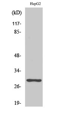
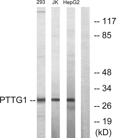
| Observed band (KD) | 30kD |
| Background | The encoded protein is a homolog of yeast securin proteins, which prevent separins from promoting sister chromatid separation. It is an anaphase-promoting complex (APC) substrate that associates with a separin until activation of the APC. The gene product has transforming activity in vitro and tumorigenic activity in vivo, and the gene is highly expressed in various tumors. The gene product contains 2 PXXP motifs, which are required for its transforming and tumorigenic activities, as well as for its stimulation of basic fibroblast growth factor expression. It also contains a destruction box (D box) that is required for its degradation by the APC. The acidic C-terminal region of the encoded protein can act as a transactivation domain. The gene product is mainly a cytosolic protein, although it partially localizes in the nucleus. Three transcript variants encoding the same protein have been fo |
| Function | developmental stage:Low level during G1 and S phases. Peaks at M phase. During anaphase, it is degraded.,disease:Has strong transforming capabilities on a variety of cell lines including NIH 3T3 fibroblasts and on athymic nude mice. Overexpressed in many patients suffering from pituitary adenomas, primary epithelial neoplasias, and esophageal cancer. No mutation in the coding sequence has been observed. The transforming capability may be due to its interaction and regulation of TP53 pathway.,domain:The N-terminal destruction box (D-box) acts as a recognition signal for degradation via the ubiquitin-proteasome pathway.,function:Regulatory protein, which plays a central role in chromosome stability, in the p53/TP53 pathway, and DNA repair. Probably acts by blocking the action of key proteins. During the mitosis, it blocks Separase/ESPL1 function, preventing the proteolysis of the cohesin c |
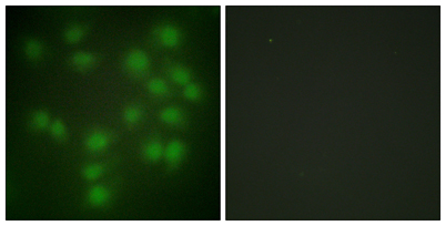
| Subcellular location | Cytoplasm. Nucleus. |
| Expression | Expressed at low level in most tissues, except in adult testis, where it is highly expressed. Overexpressed in many patients suffering from pituitary adenomas, primary epithelial neoplasias, and esophageal cancer. |

Use collapsible tabs for more detailed information that will help customers make a purchasing decision.
Ex: Shipping and return policies, size guides, and other common questions.