 Français
 English
Ribosomal Protein L34 rabbit pAb
AO-06-ES3365-100

| ELK.No | ES3365 |
| Product name | Ribosomal Protein L34 rabbit pAb |
| Reactivity | Human;Mouse;Rat |
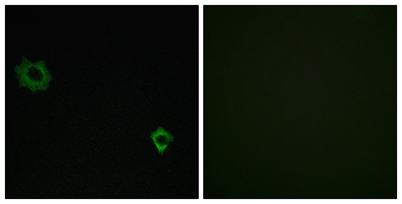
| Applications | WB;IHC;IF;ELISA |
| Other name | RPL34; 60S ribosomal protein L34 |
| Size | 100μL |
| Unit price ($) | 248 |
| Human gene ID | 6164 |
| Human Swiss-Prot | P49207 |
| Source | Rabbit |
| Isotype | IgG |
| Target | Ribosomal Protein L34 |
| Fields | >>Ribosome;>>Coronavirus disease - COVID-19 |
| Gene name | RPL34 |
| Protein name | 60S ribosomal protein L34 |
| Human gene link | View Human Gene |
| Human Swiss link | View Human Swiss-Prot |
| Mouse gene ID | |
| Mouse gene link | View Mouse Gene |
| Mouse Swiss-Prot | Q9D1R9 |
| Mouse Swiss link | View Mouse Swiss-Prot |
| Rat gene ID | |
| Rat gene link | |
| Rat Swiss-Prot | P11250 |
| Rat Swiss link | View Rat Swiss-Prot |
| Immunogen | The antiserum was produced against synthesized peptide derived from human RPL34. AA range:41-90 |
| Specificity | Ribosomal Protein L34 Polyclonal Antibody detects endogenous levels of Ribosomal Protein L34 protein. |
| Formulation | Liquid in PBS containing 50% glycerol, 0.5% BSA and 0.02% sodium azide. |
| Clonality | Polyclonal |
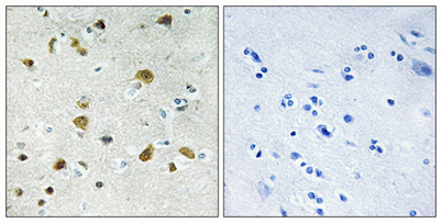
| Dilution | Western Blot: 1/500 - 1/2000. Immunohistochemistry: 1/100 - 1/300. Immunofluorescence: 1/200 - 1/1000. ELISA: 1/20000. Not yet tested in other applications. |
| Purification | The antibody was affinity-purified from rabbit antiserum by affinity-chromatography using epitope-specific immunogen. |
| Concentration | 1 mg/ml |
| Storage stability | -20°C/1 year |
| Molecular Weight (Da) | |
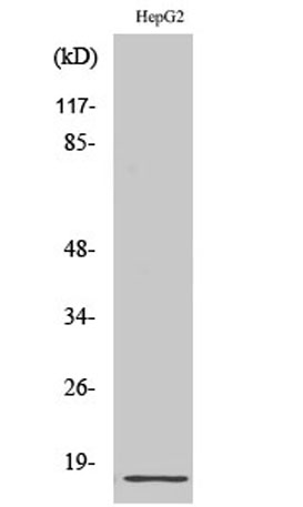
| Observed band (KD) | 13kD |
| Background | Ribosomes, the organelles that catalyze protein synthesis, consist of a small 40S subunit and a large 60S subunit. Together these subunits are composed of 4 RNA species and approximately 80 structurally distinct proteins. This gene encodes a ribosomal protein that is a component of the 60S subunit. The protein belongs to the L34E family of ribosomal proteins. It is located in the cytoplasm. This gene originally was thought to be located at 17q21, but it has been mapped to 4q. Overexpression of this gene has been observed in some cancer cells. Alternative splicing results in multiple transcript variants, all encoding the same isoform. As is typical for genes encoding ribosomal proteins, there are multiple processed pseudogenes of this gene dispersed through the genome. [provided by RefSeq, Feb 2016], |
| Function | similarity:Belongs to the ribosomal protein L34e family., |
| Subcellular location | Cytoplasm, cytosol . Cytoplasm . Endoplasmic reticulum . Detected on cytosolic polysomes (PubMed:25957688). Detected in ribosomes that are associated with the rough endoplasmic reticulum (By similarity). . |
| Expression | Bone,Epithelium,Eye,Ovary, |

Use collapsible tabs for more detailed information that will help customers make a purchasing decision.
Ex: Shipping and return policies, size guides, and other common questions.