 Français
 English

| ELK.No | ES3410 |
| Product name | SCAND1 rabbit pAb |
| Reactivity | Human;Rat;Mouse; |
| Applications | WB;IHC;IF;ELISA |
| Other name | SCAND1; SDP1; SCAN domain-containing protein 1 |
| Size | 50μL |
| Unit price ($) | 148 |
| Human gene ID | 51282 |
| Human Swiss-Prot | P57086 |
| Source | Rabbit |
| Isotype | IgG |
| Target | SCAND1 |
| Fields | |
| Gene name | SCAND1 |
| Protein name | SCAN domain-containing protein 1 |
| Human gene link | View Human Gene |
| Human Swiss link | View Human Swiss-Prot |
| Mouse gene ID | |
| Mouse gene link | |
| Mouse Swiss-Prot | |
| Mouse Swiss link | |
| Rat gene ID | |
| Rat gene link | |
| Rat Swiss-Prot | |
| Rat Swiss link | |
| Immunogen | The antiserum was produced against synthesized peptide derived from human SCAND1. AA range:61-110 |
| Specificity | SCAND1 Polyclonal Antibody detects endogenous levels of SCAND1 protein. |
| Formulation | Liquid in PBS containing 50% glycerol, 0.5% BSA and 0.02% sodium azide. |
| Clonality | Polyclonal |
| Dilution | Western Blot: 1/500 - 1/2000. Immunohistochemistry: 1/100 - 1/300. Immunofluorescence: 1/200 - 1/1000. ELISA: 1/20000. Not yet tested in other applications. |
| Purification | The antibody was affinity-purified from rabbit antiserum by affinity-chromatography using epitope-specific immunogen. |
| Concentration | 1 mg/ml |
| Storage stability | -20°C/1 year |
| Molecular Weight (Da) | |
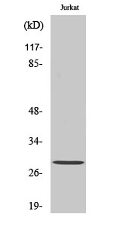
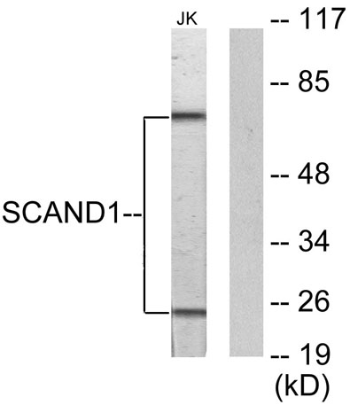
| Observed band (KD) | 20kD |
| Background | This gene encodes a SCAN box domain-containing protein. The SCAN domain is a highly conserved, leucine-rich motif of approximately 60 aa originally found within a subfamily of zinc finger proteins. This gene belongs to a family of genes that encode an isolated SCAN domain, but no zinc finger motif. This protein binds to and may regulate the function of the transcription factor myeloid zinc finger 1B. Alternate splicing results in multiple transcript variants.[provided by RefSeq, Jan 2011], |
| Function | function:May regulate transcriptional activity.,similarity:Contains 1 SCAN box domain.,subunit:Interacts with ZNF202., |
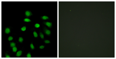
| Subcellular location | Nucleus . |
| Expression | Bone marrow,Brain,Skin, |

Use collapsible tabs for more detailed information that will help customers make a purchasing decision.
Ex: Shipping and return policies, size guides, and other common questions.