 Français
 English

| ELK.No | ES3537 |
| Product name | Syntaxin 1 rabbit pAb |
| Reactivity | Human;Mouse;Rat;Monkey |
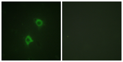
| Applications | WB;IHC;IF;ELISA |
| Other name | STX1A; STX1; Syntaxin-1A; Neuron-specific antigen HPC-1 |
| Size | 100μL |
| Unit price ($) | 248 |
| Human gene ID | 6804 |
| Human Swiss-Prot | Q16623 |
| Source | Rabbit |
| Isotype | IgG |
| Target | Syntaxin 1 |
| Fields | >>SNARE interactions in vesicular transport;>>Synaptic vesicle cycle;>>Insulin secretion;>>Huntington disease;>>Pathways of neurodegeneration - multiple diseases;>>Amphetamine addiction |
| Gene name | STX1A |
| Protein name | Syntaxin-1A |
| Human gene link | View Human Gene |
| Human Swiss link | View Human Swiss-Prot |
| Mouse gene ID | 20907 |
| Mouse gene link | View Mouse Gene |
| Mouse Swiss-Prot | O35526 |
| Mouse Swiss link | View Mouse Swiss-Prot |
| Rat gene ID | 116470 |
| Rat gene link | View Rat Gene |
| Rat Swiss-Prot | P32851 |
| Rat Swiss link | View Rat Swiss-Prot |
| Immunogen | The antiserum was produced against synthesized peptide derived from human Syntaxin 1A. AA range:1-50 |
| Specificity | Syntaxin 1 Polyclonal Antibody detects endogenous levels of Syntaxin 1 protein. |
| Formulation | Liquid in PBS containing 50% glycerol, 0.5% BSA and 0.02% sodium azide. |
| Clonality | Polyclonal |
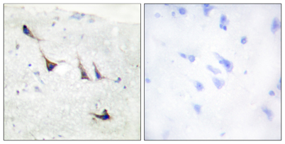
| Dilution | Western Blot: 1/500 - 1/2000. Immunohistochemistry: 1/100 - 1/300. Immunofluorescence: 1/200 - 1/1000. ELISA: 1/20000. Not yet tested in other applications. |
| Purification | The antibody was affinity-purified from rabbit antiserum by affinity-chromatography using epitope-specific immunogen. |
| Concentration | 1 mg/ml |
| Storage stability | -20°C/1 year |
| Molecular Weight (Da) | |
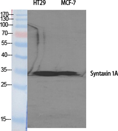
| Observed band (KD) | 35kD |
| Background | This gene encodes a member of the syntaxin superfamily. Syntaxins are nervous system-specific proteins implicated in the docking of synaptic vesicles with the presynaptic plasma membrane. Syntaxins possess a single C-terminal transmembrane domain, a SNARE [Soluble NSF (N-ethylmaleimide-sensitive fusion protein)-Attachment protein REceptor] domain (known as H3), and an N-terminal regulatory domain (Habc). Syntaxins bind synaptotagmin in a calcium-dependent fashion and interact with voltage dependent calcium and potassium channels via the C-terminal H3 domain. This gene product is a key molecule in ion channel regulation and synaptic exocytosis. Alternatively spliced transcript variants encoding different isoforms have been found for this gene.[provided by RefSeq, Sep 2009], |
| Function | disease:Haploinsufficiency of STX1A may be the cause of certain cardiovascular and musculo-skeletal abnormalities observed in Williams-Beuren syndrome (WBS), a rare developmental disorder. It is a contiguous gene deletion syndrome involving genes from chromosome band 7q11.23.,function:Potentially involved in docking of synaptic vesicles at presynaptic active zones. May play a critical role in neurotransmitter exocytosis.,similarity:Belongs to the syntaxin family.,similarity:Contains 1 t-SNARE coiled-coil homology domain.,subunit:Part of the SNARE core complex containing SNAP25, VAMP2 and STX1A. This complex binds to CPLX1. Binds SYTL4 and STXBP6. Found in a ternary complex with STX1A and SNAP25. Interacts with OTOF (By similarity). Found in a complex with VAMP8 and SNAP23. Interacts with VAPA and SYBU.,tissue specificity:Isoform 1 is highly expressed in embryonic spinal chord and ganglia |
| Subcellular location | Cytoplasmic vesicle, secretory vesicle, synaptic vesicle membrane ; Single-pass type IV membrane protein . Cell junction, synapse, synaptosome . Cell membrane . Colocalizes with KCNB1 at the cell membrane. .; [Isoform 2]: Secreted . |
| Expression | [Isoform 1]: Highly expressed in embryonic spinal cord and ganglia and in adult cerebellum and cerebral cortex. ; [Isoform 2]: Expressed in heart, liver, fat, skeletal muscle, kidney and brain. |

Use collapsible tabs for more detailed information that will help customers make a purchasing decision.
Ex: Shipping and return policies, size guides, and other common questions.