 Français
 English

| ELK.No | ES3658 |
| Product name | Tyk 2 rabbit pAb |
| Reactivity | Human;Mouse;Monkey |
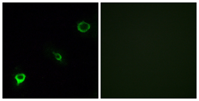
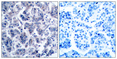
| Applications | WB;IHC;IF;ELISA |
| Other name | TYK2; Non-receptor tyrosine-protein kinase TYK2 |
| Size | 100μL |
| Unit price ($) | 248 |
| Human gene ID | 7297 |
| Human Swiss-Prot | P29597 |
| Source | Rabbit |
| Isotype | IgG |
| Target | Tyk 2 |
| Fields | >>Necroptosis;>>Osteoclast differentiation;>>NOD-like receptor signaling pathway;>>JAK-STAT signaling pathway;>>Th1 and Th2 cell differentiation;>>Th17 cell differentiation;>>Toxoplasmosis;>>Hepatitis C;>>Hepatitis B;>>Measles;>>Influenza A;>>Human papillomavirus infection;>>Kaposi sarcoma-associated herpesvirus infection;>>Herpes simplex virus 1 infection;>>Epstein-Barr virus infection;>>Coronavirus disease - COVID-19 |
| Gene name | TYK2 |
| Protein name | Non-receptor tyrosine-protein kinase TYK2 |
| Human gene link | View Human Gene |
| Human Swiss link | View Human Swiss-Prot |
| Mouse gene ID | |
| Mouse gene link | |
| Mouse Swiss-Prot | Q9R117 |
| Mouse Swiss link | View Mouse Swiss-Prot |
| Rat gene ID | |
| Rat gene link | |
| Rat Swiss-Prot | |
| Rat Swiss link | |
| Immunogen | The antiserum was produced against synthesized peptide derived from human TYK2. AA range:1020-1069 |
| Specificity | Tyk 2 Polyclonal Antibody detects endogenous levels of Tyk 2 protein. |
| Formulation | Liquid in PBS containing 50% glycerol, 0.5% BSA and 0.02% sodium azide. |
| Clonality | Polyclonal |
| Dilution | Western Blot: 1/500 - 1/2000. Immunohistochemistry: 1/100 - 1/300. Immunofluorescence: 1/200 - 1/1000. ELISA: 1/10000. Not yet tested in other applications. |
| Purification | The antibody was affinity-purified from rabbit antiserum by affinity-chromatography using epitope-specific immunogen. |
| Concentration | 1 mg/ml |
| Storage stability | -20°C/1 year |
| Molecular Weight (Da) | |
| Observed band (KD) | 134kD |
| Background | tyrosine kinase 2(TYK2) Homo sapiens This gene encodes a member of the tyrosine kinase and, more specifically, the Janus kinases (JAKs) protein families. This protein associates with the cytoplasmic domain of type I and type II cytokine receptors and promulgate cytokine signals by phosphorylating receptor subunits. It is also component of both the type I and type III interferon signaling pathways. As such, it may play a role in anti-viral immunity. A mutation in this gene has been associated with hyperimmunoglobulin E syndrome (HIES) - a primary immunodeficiency characterized by elevated serum immunoglobulin E. [provided by RefSeq, Jul 2008], |
| Function | catalytic activity:ATP + a [protein]-L-tyrosine = ADP + a [protein]-L-tyrosine phosphate.,disease:Defects in TYK2 are the cause of protein-tyrosine kinase 2 deficiency (TYK2 deficiency) [MIM:611521]; also called autosomal recessive hyper-IgE syndrome (HIES) with atypical mycobacteriosis. The syndrome consists of a primary immunodeficiency characterized by recurrent skin abscesses, pneumonia, and highly elevated serum IgE.,domain:The FERM domain mediates interaction with JAKMIP1.,function:Probably involved in intracellular signal transduction by being involved in the initiation of type I IFN signaling. Phosphorylates the interferon-alpha/beta receptor alpha chain.,online information:TYK2 mutation db,similarity:Belongs to the protein kinase superfamily. Tyr protein kinase family. JAK subfamily.,similarity:Contains 1 FERM domain.,similarity:Contains 1 protein kinase domain.,similarity:Conta |
| Subcellular location | nucleus,cytoplasm,cytosol,cytoskeleton,membrane,extrinsic component of cytoplasmic side of plasma membrane,extracellular exosome, |
| Expression | Observed in all cell lines analyzed. Expressed in a variety of lymphoid and non-lymphoid cell lines. |


Use collapsible tabs for more detailed information that will help customers make a purchasing decision.
Ex: Shipping and return policies, size guides, and other common questions.