 Français
 English
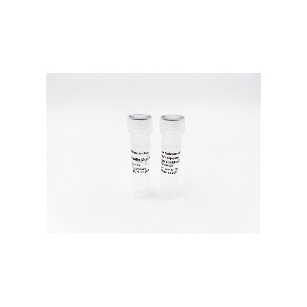

| ELK.No | ES3685 |
| Product name | Vasohibin rabbit pAb |
| Reactivity | Human;Mouse;Rat |
| Applications | WB;ELISA |
| Other name | VASH1; KIAA1036; VASH; Vasohibin-1 |
| Size | 100μL |
| Unit price ($) | 248 |
| Human gene ID | 22846 |
| Human Swiss-Prot | Q7L8A9 |
| Source | Rabbit |
| Isotype | IgG |
| Target | Vasohibin |
| Fields | |
| Gene name | VASH1 |
| Protein name | Vasohibin-1 |
| Human gene link | View Human Gene |
| Human Swiss link | View Human Swiss-Prot |
| Mouse gene ID | 238328 |
| Mouse gene link | View Mouse Gene |
| Mouse Swiss-Prot | Q8C1W1 |
| Mouse Swiss link | View Mouse Swiss-Prot |
| Rat gene ID | |
| Rat gene link | |
| Rat Swiss-Prot | |
| Rat Swiss link | |
| Immunogen | The antiserum was produced against synthesized peptide derived from human VASH1. AA range:261-310 |
| Specificity | Vasohibin Polyclonal Antibody detects endogenous levels of Vasohibin protein. |
| Formulation | Liquid in PBS containing 50% glycerol, 0.5% BSA and 0.02% sodium azide. |
| Clonality | Polyclonal |
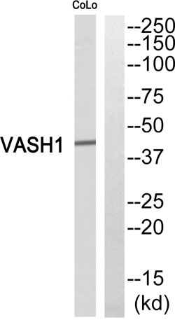
| Dilution | Western Blot: 1/500 - 1/2000. ELISA: 1/40000. Not yet tested in other applications. |
| Purification | The antibody was affinity-purified from rabbit antiserum by affinity-chromatography using epitope-specific immunogen. |
| Concentration | 1 mg/ml |
| Storage stability | -20°C/1 year |
| Molecular Weight (Da) | |
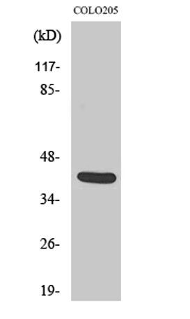
| Observed band (KD) | 40kD |
| Background | caution:Although probably secreted, it lacks a canonical signal sequence.,function:Angiogenesis inhibitor. Inhibits migration, proliferation and network formation by endothelial cells as well as angiogenesis. This inhibitory effect is selective to endothelial cells as it does not affect the migration of smooth muscle cells or fibroblasts. Does not affect the proliferation of cancer cells in vitro, but inhibits tumor growth and tumor angiogenesis. Acts in an autocrine manner. Inhibits artery neointimal formation and macrophage infiltration. Exhibits heparin-binding activity.,induction:By VEGF.,PTM:2 major forms (42 and 36 kDa) and 2 minors (32 and 27 kDa) may be processed by proteolytic cleavage. The largest form (42 kDa) seems to be secreted and the other major form (63 kDa) seems to accumulate within the cells or pericellular milieu. Polypeptide consisting of Met-77 to Arg-318 may correspond to the 27 kDa form and that consisting of Met-77 to Val-365 may correspond to the 36 kDa form.,similarity:Belongs to the vasohibin family.,tissue specificity:Preferentially expressed in endothelial cells. Highly expressed in fetal organs. Expressed in brain and placenta, and at lower level in heart and kidney. Highly detected in microvessels endothelial cells of atherosclerotic lesions., |
| Function | caution:Although probably secreted, it lacks a canonical signal sequence.,function:Angiogenesis inhibitor. Inhibits migration, proliferation and network formation by endothelial cells as well as angiogenesis. This inhibitory effect is selective to endothelial cells as it does not affect the migration of smooth muscle cells or fibroblasts. Does not affect the proliferation of cancer cells in vitro, but inhibits tumor growth and tumor angiogenesis. Acts in an autocrine manner. Inhibits artery neointimal formation and macrophage infiltration. Exhibits heparin-binding activity.,induction:By VEGF.,PTM:2 major forms (42 and 36 kDa) and 2 minors (32 and 27 kDa) may be processed by proteolytic cleavage. The largest form (42 kDa) seems to be secreted and the other major form (63 kDa) seems to accumulate within the cells or pericellular milieu. Polypeptide consisting of Met-77 to Arg-318 may corre |
| Subcellular location | Cytoplasm . Secreted . Mainly localizes in the cytoplasm (PubMed:27879017). Some fraction is secreted via a non-canonical secretion system; interaction with SVBP promotes secretion (PubMed:27879017). . |
| Expression | Preferentially expressed in endothelial cells (PubMed:15467828, PubMed:16707096). Highly expressed in fetal organs (PubMed:15467828). Expressed in brain and placenta, and at lower level in heart and kidney (PubMed:15467828). Highly detected in microvessels endothelial cells of atherosclerotic lesions (PubMed:16707096). |


Use collapsible tabs for more detailed information that will help customers make a purchasing decision.
Ex: Shipping and return policies, size guides, and other common questions.